
Page 25 - 85 cell signalling pathways
P. 25
Cell Signalling Biology Michael J. Berridge Module 2 Cell Signalling Pathways 2 25
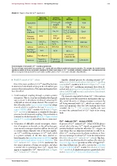
Module 2: Figure cell-specific Ca 2 + signalsomes
SKELETAL CARDIAC Ca1
MUSCLE CELL ATRIAL CELL NEURON T CELL
Receptors ET-1R/ 1R mGluR1 TCR
AngIIR M1
PLC PLC PLC PLC
Entry Ca 1.1 Ca 1.2 Ca 1.2/ Ca 2.1
V
V
channels V V Ca 2.2/ NMDAR Orai1
V
Release RYR1 RYR2 RYR2
channels InsP R2 InsP R2 InsP R1
3
3
3
PMCAs PMCA1a, PMCA1c, PMCA1a, 2a PMCA4b
1c,1d 1d,2a 3a
SERCAs SERCA1a, 1b SERCA2a SECA2b, 3 SERCA2b, 3
+
Na /Ca 2+ NCX NCX1 –
exchanger NCX1, 3
Parvalbumin
Buffers Parvalbumin Calbindin 28K
Troponin c Troponin c
Sensors Calmodulin Calmodulin
Calmodulin Calmodulin
Some examples of cell-specific Ca 2 + signalling signalsomes.
The four cell types represented here generate Ca 2 + signals with very different spatial and temporal properties. For example, the skeletal muscle
signalsome selects out those components specialized to deliver rapid pulses of Ca 2 + to activate contraction, whereas the T cell signalsome has
different components that generate the much slower repetitive pulses of Ca 2 + necessary to stimulate cell proliferation.
• NAADP control of Ca 2 + release Another classical process for releasing internal Ca 2 +
is the inositol 1,4,5-trisphosphate (InsP 3 )/Ca 2 + signalling
One of the major problems in Ca 2 + signalling has been cassette (Ca 2 + module 6 in Module 2: Figure Ca 2 + mod-
to determine how stimuli arriving at the cell surface gain ules). Other Ca 2 + -mobilizing messengers have been de-
access to these internal stores. Two main mechanisms have scribed such as sphingosine 1-phosphate (S1P), cyclic ADP
been identified: ribose (cADPR) and nicotinic acid–adenine dinucleotide
phosphate (NAADP).
1. Conformational coupling through a protein--protein The channels responsible for these Ca 2 + ON reactions
interaction. This is a very fast mechanism that depends usually have powerful inactivation mechanisms that rap-
upon a sensor in the plasma membrane interacting dir- idly curtail the entry or release processes to prevent the
ectly with an internal release channel. The receptor on cell being swamped with Ca 2 + , which can result in cell
the cell surface is the Ca V 1.1 L-type channel (a voltage
stress and apoptosis. Once the ON reactions have been
sensor), which is coupled to the type 1 ryanodine re- curtailed, the Ca 2 + OFF reactions rapidly take over
ceptor 1 (RYR1) (Ca 2 + module 4 in Module 2: Figure to return the activated level of Ca 2 + back to its resting
Ca 2 + modules). Information is transferred through a level.
process of conformational coupling. This mechanism is
restricted to skeletal muscle (Module 7: Figure skeletal
muscle E-C coupling) and perhaps also to some neur-
ons. Ca 2 + -induced Ca 2 + release (CICR)
2. Generation of diffusible second messengers. Activa- AprocessofCa 2 + -induced Ca 2 + release (CICR) plays a
tion of receptors or channels on the cell surface gen- central role in the way Ca 2 + signals are generated. This
erate second messengers that then diffuse into the cell positive-feedback mechanism whereby Ca 2 + triggers its
to activate release channels. One of the most signific- own release has two important functions in cells. It en-
ant Ca 2 + -mobilizing messengers is Ca 2 + itself, which ables Ca 2 + entering across the plasma membrane to func-
is a potent activator of the two main internal re- tion as a messenger to release Ca 2 + from the internal store
lease channels, the ryanodine receptors (RYRs) and the (Module 2: Figure Ca 2 + -induced Ca 2 + release). This func-
inositol 1,4,5-trisphosphate receptors (InsP 3 Rs).This tion of CICR was first described in cardiac cells, where the
Ca 2 + -induced Ca 2 + release (CICR) mechanism has Ca V 1.2 L-type channel provides an influx of trigger Ca 2 +
the unique property of being autocatalytic and plays that then diffuses into the cell to activate the ryanodine
a central role in generating those Ca 2 + signals that receptor 2 (RYR2) (Ca 2 + module 5 in Module 2: Figure
appear as regenerative Ca 2 + waves (Module 2: Figure Ca 2 + modules). A similar interaction is particularly evid-
Ca 2 + -induced Ca 2 + release). ent for neuronal Ca 2 + entry and release channels.
C 2012 Portland Press Limited www.cellsignallingbiology.org