
Page 64 - Noninvasive Diagnostic Techniques for the Detection of Skin Cancers
P. 64
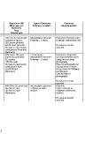
Table D-3. RCTs identified in the present technical brief
 First Author   Study   Comparisons   Setting   Population (N)   Type of Outcome,   Outcome,
 UI   Objective      Mean age, yrs    Followup duration               Adverse events
 Country      Males (%)
 year            Race
             Cancer type
 Photography
 Del Mar CB 138    To evaluate   General practitioners   Primary care   108 GPs (5784 pts with   Intermediate outcomes   Outcomes: Excision rates
 7888887   excision rate of   (GPs)  with a camera   suspicious nevus)   Followup = 2 years   of benign melanocytic nevi
 Australia   benign melanocytic   vs. GPs with no   28.2 years (pt level)
 1995   nevi   camera   43.4% male (pt level)                   No adverse events
       No data on race (study                                   reported
       conducted in Australia)
       Melanoma
 Hanrahan PF 139,140    To assess the utility  Patient received   Primary care   973 male > 50 years   Test accuracy   Outcomes: Diagnostic
 11215011   of photographs for   photographs vs.   agreed to participate   Intermediate outcomes   accuracy between those
 12370325   skin cancer   patients with no   62.0 years   Followup = 2 years   using and not using
 Australia   management   photographs   100.0% male             photography
 2000;2002   No data on race (study                             Effect of photography on
       conducted in New                                         management of lesion
       South Wales)                                             (leave lesion for followup,
       Melanoma                                                 cryotherapy)
                                                                Cost savings of
                                                                photography

                                                                No adverse events
                                                                reported
 English DR 141    To evaluate the   GPs with a camera   Primary care   468 GPs( 223 practices)  Intermediate outcomes   Outcome:
 12919990   ratio of benign to   and algorithm  vs.   setting   No data on age   Followup duration   Ratio of benign to
 Australia   malignant excision   GPs with no camera   No data on race   unclear   malignant melanoma
 2003   Melanoma                                                excised.

                                                                No adverse events
                                                                reported


 D-3