
Page 71 - Cardiac testing for coronary artery disease in potential kidney transplant recipients
P. 71
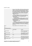
Sharma 2005  (Continued)


                                                blood pressure > 30 mm Hg, SBP rise > 230 mm Hg occurred. Patients were given an
                                                angina score: 0 = none, 1 = non-limiting angina, 2 = limiting angina. Duke score was
                                                calculated as: total treadmill time (min)-5 X magnitude of maximal ST depression
                                                (mm)- 4 X angina index. Horizontal or down sloping ST depression > 1mm measured
                                                80 ms after the J point, and ST elevation > 1 mm measured 40 ms after the J point,
                                                were regarded as positive results. The test was described as inconclusive if stopped
                                                before 85% predicted heart rate could be achieved with no cardiac symptoms or
                                                significant changes at that stage.
                                                  • DSE
                                                     ◦ An abnormal response was described as the occurrence under stress of
                                                hypokinesia, akinesia or dyskinesia in one or more resting normal segments and/or
                                                worsening of wall motion in one or more resting hypokinetic segments.
                                                  • Echocardiography
                                                  • Mitral annular calcification
                                                     ◦ The presence of mitral annular calcification was defined as an echo dense
                                                band visualised throughout systole and diastole, distinguishable from the posterior
                                                mitral valve leaflet, and located anterior and parallel to the posterior left ventricular
                                                wall on M-mode recordings.
                                                  • Resting wall motion abnormality
                                                  • Resting ECG
                                                     ◦ The ECG was considered abnormal if any of the following criteria were met
                                                in any of the standard limb leads or precordial leads, except AVR or V1: pathological Q
                                                waves, left ventricular hypertrophy by Sokolow-Lyon criteria or Cornell index, ST
                                                depression ≥ 1 mm, ST elevation ≥ 1 mm, T wave inversion or bundle branch block
                                                (QRS ≥120 ms).

                 Follow-up                      Patients were followed up for 1.32 ± 0.48 years (range 0.19 ± 2.12 years)

                 Notes

                 Table of Methodological Quality

                 Item                           Authors’ judgement             Description

                 Representative spectrum?       Yes                            ESKD patients undergoing cardiac evalua-
                 All tests                                                     tion as part of transplant workup

                 Acceptable reference standard?  Yes                           Coronary angiography with a reference
                 All tests                                                     standard threshold of ≥ 70% stenosis

                 Acceptable delay between tests?  Unclear                      Likely to be short delay between tests.
                 All tests

                 Partial verification avoided?   Yes                            All participants who underwent the index
                 All tests                                                     test received the reference standard test
                 Differential verification avoided?  Yes                        Disease status (CAD) diagnosed by coro-
                 All tests                                                     nary angiography.


                Cardiac testing for coronary artery disease in potential kidney transplant recipients (Review)  69
                Copyright © 2011 The Cochrane Collaboration. Published by John Wiley & Sons, Ltd.