
Page 5 - 65 thorax41-48_opt
P. 5
276 Laryngoscopy, Bronchoscopy, and Oesophagoscopy
ynx is seen. Secretions are sucked out and the scope is guided behind
the endotracheal tube to the cricopharyngeal inlet, where it enters the
oesophagus. Further passage of the scope is assisted by gently insufflat-
ing air to distend the lumen and by aspirating any secretions along the
way. The scope is passed all the way down into the stomach; Then, on
withdrawal, a careful note is made of any pathology that has been noted
previously. Any procedures that need to be carried out are then done
with the scope positioned at the appropriate site. Many devices, such
as forceps, needles, and electrosurgical knives, among others, available
for therapeutic and diagnostic purposes can be inserted through an
instrument channel (Figure 41.10).
Dilatation of strictures (Figures 41.11 and 41.12) can be done either
with the direct endoscopic view or with radiological screening. For
this purpose, contrast is used to fill the balloon, and the procedure is
observed on the x-ray screen. Figure 41.11: Oesophageal balloon (left) for dilatation of stricture (right) – accurate
placement of the balloon and monitoring of the pressure used are essential.
Complications
Complications are rare and include minor haemorrhage, injury to the
larynx and hypopharynx and infections. Perforation can occur espe-
cially following deep biopsy, forceful dilation of strictures, or during
removal of foreign bodies.
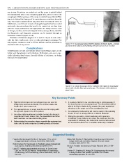
Figure 41.12: Direct endoscopic view to a stricture after repair of oesophageal
atresia with a flexible fibre-optic gastroscope. The diameter of this stricture is
about 3 mm.
Key Summary Points
1. Rigid bronchoscopes and oesophagoscopes are used for 5. It is always helpful to use a laryngoscope to guide passage of
foreign body removal and biopsy. For all other cases, use the bronchoscope or oesophagoscope. The anaesthetist will be
flexible fibre-optic instruments. able to do this to display to the surgeon the exact anatomical
structures at the upper end of the aerodigestive tract.
2. The appropriate size of scope must be used in keeping with
the age and physical size of the patient. 6. An emergency tracheostomy set must be ready in the operating
theatre in case of airway problems or difficult intubation.
3. Use video systems whenever possible because the image is
magnified and much clearer. Also, the anaesthetist and other 7. During the procedure, careful monitoring of the patient is
team members can see what is being done. mandatory. If any problems are noted, the endoscopy must be
suspended. If necessary, the instruments must be removed and
4. Before starting the procedure, always assemble the equipment and the anaesthetist allowed to re-establish satisfactory ventilation.
make sure that every component is working exactly as intended.
Suggested Reading
Edwards MJ, Greenland KB, Allen P, Cumpston P. The correct Mathur NN, Pradhan T. Rigid pediatric bronchoscopy for bronchial
laryngoscope blade for the job. Anaesthesia 2009; 64:95. foreign bodies with and without Hopkins telescope. Indian
Pediatrics 2003; 40:761–765.
Holm-Knudsen RJ, Rasmussen LS. Paediatric airway management:
basic aspects. Acta Anaesthesiol Scand 2009; 53:1–9. Nicolai T. Pediatric bronchoscopy. Pediatr Pulmonol 2001; 31:150–
164.
Lobe TE. Pediatric gastrointestinal endoscopy. In: Scott-Conner
CEH, ed. The SAGES Manual: Fundamentals of Laparoscopy, Shinhar SY, Strabbing RJ, Madgy DN. Esophagoscopy for removal
Thoracoscopy, and GI Endoscopy, 2nd ed. Birkhauser, 2005, Pp of foreign bodies in the pediatric population. Int J Pediatr
747–751. Otorhinolaryngol 2003; 67: 977–979.