
Page 4 - 65 thorax41-48_opt
P. 4
Laryngoscopy, Bronchoscopy, and Oesophagoscopy 275
Trauma to lips, teeth, epiglottis, and larynx with subsequent oesophagus, the neck is extended by placing a roll under the shoulders.
airway oedema, especially subglottic oedema, are complications This brings the axis of the scope into a straight line with the oesophagus
associated mainly with rigid bronchoscopes. If stridor is present in and it is advanced under direct vision to the cardia. It is sometimes
recovery, nebulised epinephrine should be administered. Intravenous helpful to use a 0-degree telescope to view the distal lumen as the
administered dexamethasone also produces relief of stridor but takes 1 scope is advanced. In this case, the videocamera can be attached to the
to 2 hours to act. eyepiece to provide an image on the monitor (Figure 41.9).
Damage to the tracheobronchial tree with pneumothorax or Flexible endoscopes
pneumomediastinum is rare. Pneumothorax or pneumomediastinum Currently, most of the diagnostic and therapeutic procedures are per-
can also be the consequence of air trapping, as passive expiration formed with flexible endoscopes. Although it is possible to insert the
cannot overcome the resistance in the airway obstructed by the flexible scope in an awake patient under sedation, most children will
instrument. Air trapping can also lead to diminished venous return and require general anaesthesia for this procedure. The patient is gener-
reduced cardiac output. ally placed in the lateral position lying on the left side, although some
Local anaesthetic overdose may cause serious bradycardia and even death. surgeons prefer the supine position. The tip of the scope is angulated
Infections are a problem, especially in flexible bronchoscopy. A into a curve to follow the back of the tongue. On insertion, the phar-
major problem is proper disinfection of the suction channel and valves.
Leak detection should be performed regularly because bacteria may
penetrate into fissures around the optic fibres and cables.
Haemorrhage from granulations or haemangiomas is usually a
minor problem and settles spontaneously.
Oesophagoscopy
In the earliest endoscopic procedures to visualise the oesophagus, only
rigid instruments were available. These instruments are similar to rigid
bronchoscopes except they lack side holes at the distal end and the
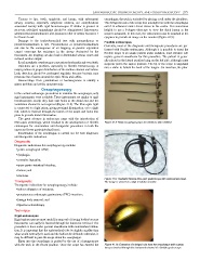
ventilation channel is not required (Figure 41.8). The fibre-optic light
is connected to a light prism, giving proximal illumination, or to a light
rod, which is inserted through the lumen of the scope and locks into
place to provide distal illumination.
The great advance in endoscopy came with the introduction of
fibre-optic technology, which resulted in the development of flexible Figure 41.8: Rigid oesophagoscopes for infants to older children.
endoscopes for examination and therapeutic procedures in both the
upper and lower gastrointestinal tracts.
Examination of the oesophagus is carried out for both diagnostic
and therapeutic indications.
Diagnostic
Diagnostic indications for oesophagoscopy include:
• gastro-oesophageal reflux;
• dysphagia;
• corrosive ingestion;
• upper gastro-intestinal bleeding;
• trauma; and
• strictures.
Figure 41.9: Paediatric flexible fibre-optic gastroscope with videocamera head.
Therapeutic The image is viewed on a high-resolution monitor.
Therapeutic indications for oesophagoscopy include:
• balloon dilatation of strictures;
• percutaneous endoscopic gastrostomy (PEG) insertion;
• foreign body removal; and
• injection sclerotherapy.
Technique
Rigid endoscopes
Rigid endoscopes are most useful for removal of foreign bodies because
instruments can easily be inserted through the lumen for retrieval. The
procedure is done under general anaesthesia with endotracheal intuba-
tion. It is important that the endotracheal tube be slightly smaller than
what would normally be used and the balloon be deflated; otherwise, it
may be difficult to pass the scope down the oesophagus.
Entry into the oesophagus is guided by the use of a laryngoscope
with the neck in the flexed position. Once the scope has entered the Figure 41.10: Extraction of a lodged coin from the oesophagus with a grasp
forceps inserted through the instrument channel of a flexible gastroscope