
Page 3 - 65 thorax41-48_opt
P. 3
274 Laryngoscopy, Bronchoscopy, and Oesophagoscopy
collapse and consolidation distal to the obstruction, but it may also be
without pathologic findings.
Indication for bronchoscopy is a positive history and clinical signs
of aspiration.
Usually a rigid technique is used for the removal of foreign bodies.
With the patient deeply anaesthetised, a rigid ventilation bronchoscope
is introduced under direct view into the trachea. The foreign body is
extracted with special grasping forceps. In most cases, the foreign body is
too large to be removed through the bronchoscope, so the object, forceps,
and the bronchoscope have to be removed together as a single unit.
Great danger ensues when the foreign body is lost in the trachea or
in the subglottic space obstructing the airway. If the object cannot be
removed quickly, it should be pushed down into a main stem bronchus
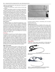
to allow oxygenation. Then a second attempt at removal can be made. Figure 41.5: Storz ventilation bronchoscope with Hopkins rod telescope. A
After removal of the foreign body, the presence of a second foreign battery-powered light source is connected to the telescope.
body should be excluded.
Airway stenosis by using an apnoeic technique or alternatively with the newborn
An important indication for interventional bronchoscopy is treat- breathing spontaneously.
ment of airway stenoses. Laser therapy of subglottic haemangiomas Flexible bronchoscopes
is favoured by some, whereas others use application of intralesional
The flexible fiberscope consists of a flexible tube that contains a fibre-
steroids followed by intubation. Subglottic granulation tissue and viral
optic system that transmits an image from the tip of the instrument to
papillomas can be treated with the intralesional injection of drugs (cor-
an eyepiece (Figures 41.6 and 41.7). Another technical advance is the
ticosteroids and chemotherapeutic agents). Subglottic cysts, which can
video scope. In these instruments, a video chip positioned at the tip of
develop after intubation, can be resected with a laser or with special
the bronchoscope replaces the glass fibre bundle. This design avoids the
forceps. These interventions require the availability of an intensive care
inherent susceptibility of a fibre bundle to damage. Digital processing
unit because many children need to remain intubated due to secondary of the image is also possible. Using Bowden cables connected to a lever
swelling of the subglottic area.
at the handpiece, the tip of the instrument can be oriented, allowing the
Technique practitioner to navigate the instrument into individual lobe or segment
In many parts of the world, the use of flexible endoscopes for diagnos- bronchi. Small fibre-optic endoscopes down to 2.2 mm in external
tic purposes is regarded as a standard, but in many other locations, the diameter are available, but these very small instruments lack a chan-
availability and cost of flexible bronchoscopes limit the use of these nel for suctioning and instrumentation. The fiberscope can be inserted
expensive and fragile instruments. Adequate assessment of the supra- through the nose or the mouth under local anaesthesia with or without
glottis, subglottis, and the trachea is possible in most cases, however, sedation. Very young children often need deep sedation or anaesthesia.
by using a telescopic rod alone with the patient breathing spontaneously Otherwise, only suboptimal information can be obtained due to move-
with 100% oxygen and a volatile agent, usually halothane or sevoflu- ment, coughing, and obstructed view.
rane. Rigid endoscopy is ideal for therapeutic interventions such as for- Complications
eign body extraction or laser surgery. The main disadvantage of the rigid Complications include hypoxia, hypoventilation, and hypercapnia for
technique is that it can be used only under anaesthesia, whereas flexible many reasons, including obstruction of the airway or deep sedation.
bronchoscopes can be used under sedation and local anaesthesia.
Rigid bronchoscopes
Rigid ventilation bronchoscopes consist of a light metal tube. A port
at the distal end allows the attachment of an anaesthetic T-piece for
ventilation. Light is transmitted over a prism at the distal end of the
tube. The ventilation scope can be used with spontaneous or controlled
breathing. The scope can be used with the Hopkins rod telescope
for diagnostic procedures (Figure 41.5). With the telescope in place,
ventilation and examination are possible under excellent visual condi-
tions. However, the telescope narrows the lumen of the bronchoscope, Figure 41.6: Small flexible fibreoptic bronchoscope with suction/irrigation and
increasing airflow resistance and making breathing difficult. This is biopsy channel.
particular a problem with the smallest bronchoscopes. For therapeutic
procedures the ventilation bronchoscope is used with special equip-
ment, such as grasping forceps, for extraction of foreign bodies.
The Hopkins rod telescope is an endoscopic telescope in which
the air-containing spaces between the conventional series of lenses
are replaced with glass rods with polished ends separated by small air
lenses. This system transmits more light, yields greater magnification,
and provides greater depth and breadth of field than conventional lens
systems. The instrument is inserted under direct laryngoscopy with a
standard laryngoscope through the mouth under general anaesthesia,
with the patient lying in a supine position. The smallest available
telescope has a diameter of less than 2 mm. With this instrument,
diagnostic bronchoscopy is possible even in very small newborns. Figure 41.7: Standard flexible fibre-optic bronchoscope with full deflection,
suction/irrigation channel, and biopsy channel for instruments.
In this case the Hopkins rod telescope alone can be inserted either