
Page 2 - 65 thorax41-48_opt
P. 2
Laryngoscopy, Bronchoscopy, and Oesophagoscopy 273
bronchoscope has no suction or instrument channel and is mostly used
by anaesthetists for intubation in difficult head and neck cases. The
standard bronchoscope has an instrument/suction channel and can be
used for therapeutic indications, although the rigid instrument is greatly
superior in this respect. Both the standard flexible bronchoscope and
the nasopharyngoscope are used to evaluate laryngomalacia and vocal
cord paralysis.
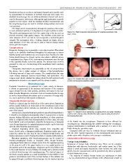
The image can be viewed directly through the eyepiece of the scope;
in more advanced systems, it is displayed on a high-resolution monitor. Figure 41.2: Rigid suspension laryngoscope for surgical procedures in the
The newer nasolaryngoscopes have the camera chip at the tip and can upper airway
provide extremely high quality images. The ultrafine scopes have an
outer diameter of 2.2 mm but do not incorporate a suction/irrigation
channel. The instruments with a working channel are larger, with an
outer diameter of 4.9 mm, and can be used to remove foreign bodies
and to perform biopsies.
Complications
Diagnostic laryngoscopy is generally a very safe procedure. The patient
needs to be carefully monitored throughout the endoscopy to ensure
that the airway and ventilation are not compromised. Facilities for intu-
bation should always be at hand, and in cases where a difficult airway
is anticipated (see Figure 41.4), tracheostomy instruments must be kept
in the operating theatre next to the patient. The surgical team must be
prepared to carry out a tracheostomy if the anaesthetist fails to intubate
the patient.
Therapeutic interventions are potentially at risk of compromising
the airway, due to either oedema or collapse of the larynx/trachea
following removal of large neck masses. The complications that may
occur include laryngeal oedema, haemorrhage, and perforation. The
surgeon must decide whether the patient should be left intubated with Figure 41.3: Flexible fibre-optic nasopharyngoscope (left) showing normal view
postoperative intensive care support until the airway is stable. (centre) and laryngeal papilloma (right).
Bronchoscopy
Paediatric bronchoscopy is indicated for a wide variety of diseases.
It allows an assessment of the anatomy and function of the complete
upper airway from the nasal passage, pharynx, and larynx to the seg-
ment bronchi. Diagnostic procedures such as bronchoalveolar lavage,
as well as interventional procedures such as extraction of foreign bod-
ies, can be performed with special instruments.
Diagnostic Bronchoscopy
Stridor is a clinical sign for obstruction of the upper airway. Inspiratory
stridor usually indicates an obstruction of the extrathoracic part of the
airway. Expiratory stridor indicates an obstruction of the intrathoracic
part of the airway.
In most cases, congenital inspiratory stridor is caused by laryngomalacia. Figure 41.4: Massive tumour (teratoma) occupying whole oral cavity. Intubation
could be done only by using a flexible fibre-optic scope to guide placement of
It should be investigated endoscopically when it is progressive or causes the endotracheal tube.
apnoea, feeding difficulties and growth retardation, or when symptoms
point to a diagnosis other than laryngomalacia. In these cases, one may
find bilateral vocal cord paralysis, subglottic hemangioma, or laryngeal of the fistula into the oesophagus. Diagnosis is then affirmed by
cysts. Proper diagnosis of congenital inspiratory stridor can be done only oesophagoscopy, demonstrating the catheter entering the oesophagus.
with the child breathing spontaneously. Surgical identification is facilitated with a catheter or a wire in the
Acquired inspiratory stridor may originate from subglottic scar fistula during the operative repair.
tissue, ductal cysts, or laryngeal papillomas. Expiratory stridor may be Laryngeal clefts are easy to overlook because redundant mucosa
caused by asthma but also may be due to inhaled foreign bodies and fills the cleft. Careful inspection of the interarytenoid and posterior
tracheomalacia as a result of tracheobronchial or vascular malformations. glottis region with a Hopkins rod telescope is mandatory.
Recurrent aspiration with bronchopneumonias can be caused by Interventional Bronchoscopy
broncho-oesophageal fistulas or laryngeal clefts. H-type broncho- Foreign body inhalation
oesophageal fistula takes an oblique course from the cephalad opening
on the posterior wall of the upper trachea to a more caudal position on Symptoms of foreign body inhalation vary. There can be complete
the anterior wall of the oesophagus. Diagnosis can be very difficult due obstruction with hypoxia, bradycardia, and cardiac arrest, but if the
to the small diameter of some fistulas, but usually can be achieved with object is small and passes beyond the main bronchi, the child may
combined bronchoscopy and oesophagoscopy. The tracheal aspect of quickly become asymptomatic and be presented only when symptoms
the fistula usually appears as a small prominence in the midline of the of distal obstruction occur.
posterior membranous wall of the cervical trachea. A fine catheter can The majority of inhaled foreign bodies are radiolucent. A chest
be passed through a ventilation bronchoscope into the tracheal opening x-ray may show unilateral hyperinflation of the affected side as well as