
Page 14 - 65 thorax41-48_opt
P. 14
Tracheomalacia 285
Innominate vein
Three sutures in
Ascending the ascending aorta
aorta
Ascending
Pericardium aorta
opened
SVC
Right atrial
appendage Heart
(A) Pulmonary (B)
artery
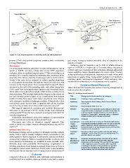
Figure 43.2: (A) Surgical approach to ascending aorta; (B) suture placement.
pressure (CPAP) may provide temporary assistance but is not suitable geal atresia. Aortopexy leads to immediate relief of symptoms in the
for long-term therapy. majority of infants.
Aortopexy Aortopexy may be required in up to 10% of infants following
For severe tracheomalacia, especially for cases complicated by “dying repair of OA/TOF, at a median age of 7 months. Ninety-five percent
spells” or ALTEs, and those infants who remain CPAP dependent, of these cases have resolution of their symptoms, although almost half
6
aortopexy offers an excellent surgical option. 6,7,8 The crucial step in an require antireflux surgery (fundoplication) for severe reflux. Overall,
aortopexy is to ventrally suspend the ascending aorta, suturing it to the aortopexy affords good symptomatic improvement in such infants, with
underside of the sternum, thereby creating space anterior to the trachea. indications for surgery being “dying spells”, inability to be extubated,
14
Access to the aortic arch is achieved via either a median sternotomy expiratory stridor, and recurrent pneumonia. When aortopexy fails,
or a left anterior thoracotomy (through the bed of the third rib), with insertion of an airway stent or a tracheostomy may be required.
resection of the thymus gland. Three nonabsorbable Prolene™ sutures Evidence-Based Research
are placed in the wall of the ascending aorta, each suture taking bites Tables 43.4 and 43.5 present case reviews involving management of
of the vessel from its intrapericardial segment to the innominate artery. tracheomalacia by aortoplexy.
These sutures can be passed through the infant sternum or sutured to its
deep periosteum. The assistant depresses the sternum as the sutures are Table 43.4: Evidence-based research.
tied with minimal tension (Figure 43.2). Complications from surgery Title Management of tracheomalacia by aortopexy
include bleeding from major vessel injury and phrenic nerve damage Authors E M Kiely, L Spitz, and R Brereton
with subsequent ipsilateral diaphragm paralysis. Alternatively, a low Institution The Hospital for Sick Children, Great Ormond Street,
cervical skin crease incision with a manubrial split affords excellent London, UK
access for surgery under direct vision, with improved cosmesis. 9 Reference Pediatr Surg Int 1987; 2:13–15
The surgical approach to aortopexy now includes thoracoscopy, Problem The problem is symptomatic tracheomalacia in infants with
with repair of the primary OA/TOF having already been undertaken congenital tracheo-oesophageal anomalies. Indications for
endoscopically. It has also been employed in aortopexy undertaken for surgery included respiratory distress, recurrent apnoea,
10
vascular compression. 11 cyanosis or “dying spells”, worsening stridor, or repeated
hospital admissions for respiratory infections
In specialist cardiothoracic units, short segments of tracheomalacia
may be resected and a primary anastomosis performed. Intervention Aortopexy
Glossopexy may offer an alternative surgical approach. This Comparison/ Case review (level 4). A review of 210 infants with tracheo-
serves to anchor the tongue forward, although aortopexy may still be control oesophageal anomalies admitted over a six and a half year
period. Twenty-five infants underwent an aortopexy, 22
required. 13 (quality of having had repair of an oesophageal atresia and three who
evidence)
Endoluminal Stenting had primary tracheomalacia.
Endoluminal stenting appears an attractive treatment modality, initially Outcome/ Seventeen infants had immediate and dramatic relief of
symptoms, and the other five were greatly improved. The
arising from a need to manage malignant airway compromise in the effect operation failed in one patient.
adult population. Technology used in endovascular stenting has further
advanced the techniques. Balloon-expandable metallic or silicone-type Historical Aortopexy had previously been described as a surgical
option for the treatment of symptomatic vascular
stents placed at bronchoscopy are available in some specialist units. significance/ compression of the trachea. This was the first description
comments
However, they carry potentially life-threatening complications of of this surgical procedure for patients with congenital
bleeding, granulation tissue formation, luminal obstruction, and erosion oesophageal anomalies. It demonstrated an excellent
into adjacent blood vessels. Removal of these stents is also hazardous outcome from aortopexy for children with significant
tracheomalacia, and recommended early surgery.
but they can offer an alternate mode of management in selected cases.
12
Outcome
Long-term follow-up of children with significant tracheomalacia is
mainly derived from studying infants previously treated with oesopha-