
Page 13 - 65 thorax41-48_opt
P. 13
284 Tracheomalacia
Table 43.2: Signs and symptoms of tracheomalacia.
Widely patent
Mild Harsh, barking TOF cough lumen
Exacerbating Crying
events Coughing
Feeding (especially food bolus)
Acute distress
Moderate Expiratory stridor Normal
Wheeze Narrow lumen
Chronic cough
Recurrent respiratory infections
Feeding difficulties
Failure to thrive
Respiratory distress (tachypnoea, intercostal recession,
hypoxia) Severe: “fish mouth”
Severe Severe hypoxia
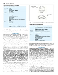
Figure 43.1: Airway in cross section showing varying degrees of tracheomalacia.
Biphasic stridor
Cyanosis
Table 43.3: Management of tracheomalacia.
Reflex apnoea (vagal stimulation)
“Dying spells” or acute life-threatening events, which may Supportive Frequent, small oral feeds
be fatal. NGT feeding (during times of acute respiratory infections)
CPAP
Intubation and positive pressure ventilation
rigid and afford better support of the airway. However, it may take
many years for a TOF cough to disappear, and for some this clinical Co-morbid Antibiotics for acute pneumonia
sign will persist into adult life. pathology Supplemental oxygen (pneumonia, RSV infections)
Assessment Antireflux therapy for GOR
Surgical Aortopexy
The need for investigation should be guided by the severity of symp-
Tracheostomy
toms demonstrated by the child. For infants who have already had
Correction of vascular rings or extrinsic compression
surgical correction for OA, a high index of suspicion should alert the
(vascular, mediastinal mass)
clinician to signs of developing tracheomalacia. Close observation and
Endobronchial stenting
timely investigation are recommended. For older children presenting
Glossopexy
with significant tracheomalacia, vascular or mediastinal compression
should be considered. Other conditions that may cause diagnostic Antireflux surgery (fundoplication)
confusion, including a laryngeal cleft, laryngomalacia and H-type TOF
(H-TOF), should be excluded or confirmed by laryngobronchoscopy.
abnormal vascular anatomy or a mediastinal mass. These methods are
A plain chest x-ray is of limited diagnostic value, although it
less helpful in identifying tracheomalacia, which is a dynamic process.
may show a mediastinal mass. A lateral chest x-ray may demonstrate
Vascular anomalies may require further specialist investigations.
localised narrowing of the trachea. Flow volume loops are able to
demonstrate major airway compromise, but the impracticalities of Management
performing them in babies and infants limit their use except in specialist The management of tracheomalacia is summarised in Table 43.3.
research facilities. Treatment is initially focused on managing predisposing conditions. In
A bronchoscopy performed under general anaesthetic is the initial cases of compression from a vascular ring or aberrant vessel, surgical
investigation of choice. This will both establish the diagnosis and correction may be required. This should be performed by a paediat-
assess the degree and location of any airway collapse. It is important ric cardiothoracic surgeon and is tailored to the underlying vascular
to ensure that the child continues to breathe spontaneously and does anomaly. Most commonly, surgical correction involves division of the
not receive intravenous muscle relaxation. A rigid bronchoscopy will smaller arch in cases of a double aortic arch, division of the ligamen-
allow visualisation of the supraglottic, laryngeal, and tracheobronchial tum arteriosum when seen with other vascular rings, or reimplanting an
tree. Flexible bronchoscopy, ideally via a laryngeal mask, provides aberrant vessel (typically the pulmonary artery in cases of a PA sling).
superior assessment of any airway collapse. The AP diameter of the However, tracheomalacia may persist or progress following correction
airway reduces during expiration, and in severe cases, the anterior and of an underlying pathology, such as a OA/TOF.
posterior tracheal walls will touch and occlude the airway entirely. Not all children will require intervention, especially when symptoms
The site of collapse is confirmed by a typical “fish mouth” appearance are mild. Appropriate medical treatment for GOR is started, and, when
(Figure 43.1). necessary, antireflux surgery may be undertaken. Respiratory infections
An upper gastrointestinal (UGI) contrast study with both AP and require appropriate antibiotic therapy. RSV infections often require
lateral views of the entire oesophagus is recommended. This can clearly hospital admission and even respiratory support in the acute phase.
suggest a vascular ring and may demonstrate GOR. A double aortic Oral feeding may be problematic during this time, and nasogastric tube
arch is suggested by both a right and left lateral indentation of the (NGT) supplementation may be required.
oesophageal outline seen in the AP view and a posterior indentation on As the degree of tracheomalacia increases, conservative measures
the lateral view. This differs from the normal left-sided indentation by will not suffice. Supplemental oxygen may be required and should be
the normal aortic arch. available at home. The parents should receive resuscitation training.
Cross-sectional imaging of the chest with computed tomography Adjustment of oral dietary regimens and periods of NGT feeding may
(CT) or magnetic resonance imaging (MRI) will identify either be required. Support of the airway with continuous positive airway