
Page 83 - 85 cell signalling pathways
P. 83
Cell Signalling Biology Michael J. Berridge Module 2 Cell Signalling Pathways 2 83
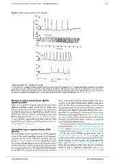
Module 2: Figure thimerosal-induced Ca 2 + signalling
Thimerosal-induced Ca 2 + oscillations in oocytes.
A.The normal Ca 2 + oscillation induced by sperm fusion in a mouse oocyte. B--D. Examples of Ca 2 + oscillations induced by addition of the oxidizing
agent thimerosal at three different concentrations: B, 100 μM; C,10 μM; D,1 μM. Reproduced from Cheek, T.R., McGuinness, O.M., Vincent, C.,
Moreton, R.B., Berridge, M.J. and Johnson, M.H. (1993) Fertilisation and thimerosal stimulate similar calcium spiking patterns in mouse oocytes but
by separate mechanisms. Development 119, 179--189, with permission from The Company of Biologists; see Cheek et al. 1993.
Mitogen-activated protein kinase (MAPK) Raf-1, A-Raf and B-Raf that phosphorylate two serine
signalling toolkit residues on the MAPK/ERK kinase (MEK) components
There is an extensive mitogen-activated protein kinase MEK1/2. The latter are dual-specificity protein kinases
(MAPK) signalling toolkit, which can be divided into that phosphorylate the tyrosine and threonine residues of
different functional components such as the transducers, the characteristic MAPK components ERK1/2 that are re-
the MAPK kinase kinases (MAPKKKs), MAPK kinases sponsible for stimulating the downstream effectors, many
(MAPKKs), MAPKs, MAPK scaffolding proteins and of which are transcription factors (Module 2: Figure ERK
MAPK target proteins (Module 2: Table MAPK signalling signalling). There is thus a linear transfer of information
toolkit). Specific components from this toolkit are then through a phospho-relay system based on a sequential
assembled into the different signalling pathways (Module series of phosphorylation events.
2: Figure MAPK signalling). An important feature of this ERK pathway, which can be
activated by both protein tyrosine kinase-linked receptors
(PTKRs) and by G protein-coupled receptors (GPCRs),is
Extracellular-signal-regulated kinase (ERK)
pathway its spatial organization (Module 2: Figure ERK signalling).
The extracellular-signal-regulated kinase (ERK) pathway In the case of the PTKRs, growth factors such as
is one of the major signalling cassettes of the mitogen- platelet-derived growth factor (PDGF) usually cause
activated protein kinase (MAPK) signalling pathway receptor dimerization, which allows the cytosolic tyrosine
(Module 2: Figure MAPK signalling). It performs a num- kinase domains to come together and to phosphorylate
ber of important signalling functions, including the control each other (Module 1: Figure PDGFR activation).
of cell proliferation and the synaptic plasticity responsible These phosphorylated residues then function as docking
for learning and memory. The main MAPK/ERK kinase motifs to pull in signalling components such as Shc,
kinase (MEKK) components are the Raf family members growth factor receptor-bound protein 2 (Grb2) and
C 2012 Portland Press Limited www.cellsignallingbiology.org