
Page 84 - 85 cell signalling pathways
P. 84
Cell Signalling Biology Michael J. Berridge Module 2 Cell Signalling Pathways 2 84
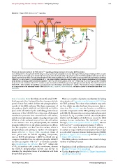
Module 2: Figure ROS effects on Ca 2 + signalling
Antigen
BCR
PtdIns4,5P
2 DUOX
O 2
P P
P BLNK P PLC 2
P
P P P P IP
Lyn P 3 H O
2 2
P Syk 2+ +
P Btk Ca
SHP1
SHP1
SHP1
_
_
_
Reciprocal interaction between the ROS and Ca 2 + signalling pathways during B cell receptor (BCR) activation.
Cross-linking the B cell receptor (BCR) with antigen sets up a protein phosphorylation cascade that begins with Lyn phosphorylating both the receptor
and Syk. The latter then binds to the phosphorylated receptor, where it phosphorylates the adaptor protein B cell linker (BLNK) and the tyrosine kinase
Bruton’s tyrosine kinase (Btk). The latter phosphorylates BLNK and phospholipase Cγ2(PLCγ2), which associates with BLNK and begins to hydrolyse
PtdIns4,5P 2 to form inositol 1,4,5-trisphosphate (IP 3 ). This phosphorylation cascade is kept in check by the tyrosine phosphatase Src homology 2
(SH2) domain-containing protein tyrosine phosphatase-1 (SHP-1). One of the functions of the Ca 2 + released by IP 3 is to set up a positive-feedback
loop based on the activation of the Ca 2 + -sensitive enzyme dual oxidase (DUOX) that generates hydrogen peroxide (H 2 O 2 ). The latter feeds back to
inhibit SHP-1, which enables the signalling cascade to work more effectively in generating Ca 2 + signals. This formation of H 2 O 2 is highly localized
as a microdomain in the immediate vicinity of the BCR (Module 2: Figure ROS microdomains). This figure is based on information taken from Singh
et al. 2005.
Son-of-sevenless (SoS) that then activate the small GTP- There are a number of putative mechanisms for linking
binding protein Ras. Activated Ras then interacts with the the activation of G protein-coupled receptors (GPCRs) to
protein kinase Raf, which initiates the phosphorylation the ERK pathway. The release of βγ subunits may activ-
cascade of the ERK pathway. The three components of ate Src, which can then feed into the processes that ac-
this pathway (Raf-1, MEK1/2 and ERK1/2) are held in tivate Ras. The arrestins can also function as scaffolds to
place at the cell surface by the scaffolding protein kinase assemble components of the ERK pathway such as Raf1
suppressor of Ras 1 (KSR1). Up to this point, all the signal and ERK1/2. Alternatively, activation of phosphoinositide
transduction processes have occurred at the cell surface, hydrolysis by G q to produce inositol 1,4,5-trisphosphate
and the next information transfer step depends upon the (InsP 3 ) and diacylglycerol (DAG) can access the ERK
diffusion of the activated enzyme from the cell surface pathway via two mechanisms. The InsP 3 /Ca 2 + can act
to the nucleus. Once it is phosphorylated, the activated through proline-rich tyrosine kinase 2 (Pyk2), whereas
phospho-ERK1/2 leaves the plasma membrane to diffuse DAG and Ca 2 + act through protein kinase C (PKC).
into the cytoplasm and then into the nucleus, where it One of the major functions of the ERK pathway is
phosphorylates and activates a number of transcription to activate a range of different transcription factors such
factors (Module 2: Figure ERK signalling). Some of as cyclic AMP response element-binding protein (CREB)
these genes code for MAPK signalling components, (Module 4: Figure CREB activation) and Elk-1 (Module
such as MAPK phosphatase 1 (MKP-1), which sets up 4: Figure ETS activation).
a negative-feedback loop. In addition, phospho-ERK1/2 This ERK pathway contributes to the control of a large
can also act together with Ca 2 + to stimulate cytoplas- number of cellular processes:
mic phospholipase A 2 (cPLA 2 ).The Ca 2 + induces the
cPLA 2 to associate with cytosolic membranes, whereas • Regulation of cell proliferation such as T cell activation
the ERK1/2 phosphorylates Ser-505 and Ser-727 to (Module 9: Figure TCR signalling)
stimulate the enzymatic release of arachidonic acid from • Cardiac hypertrophy (Module 12: Figure hypertrophy
phospholipid precursors. signalling mechanisms)
C 2012 Portland Press Limited www.cellsignallingbiology.org