
Page 85 - 85 cell signalling pathways
P. 85
Cell Signalling Biology Michael J. Berridge Module 2 Cell Signalling Pathways 2 85
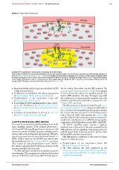
Module 2: Figure ROS microdomains
BCR
Resting
SHP1 DUOX SHP1
Lyn SHP1
SHP1 SHP1 SHP1 SHP1
SHP1 Syk BLNK PLC IP 3
SHP1 Btk SHP1
SHP1 2+ SHP1
Ca
SHP1
SHP1
SHP1 SHP1 SHP1
SHP1 SHP1
SHP1 SHP1 SHP1
SHP1
Antigen
Stimulated
DUOX SHP1
Lyn SHP1 SHP1
SHP1 SHP1 SHP1 SHP1
SHP1 BLNK PLC IP
SHP1 Syk 3 SHP1
SHP1 Btk SHP1 SHP1
SHP1 2+
SHP1 H O microdomain Ca
2 2
SHP1 SHP1
SHP1 SHP1 SHP1 SHP1 SHP1
SHP1 SHP1 SHP1
PROLIFERATION
Localized ROS signalling in a microdomain surrounding the B cell receptor.
Under resting conditions, the protein phosphorylation cascade that operates between Lyn, Syk, Bruton’s tyrosine kinase (Btk) and phospholipase C
(PLC) (Module 2: Figure ROS effects on Ca 2 + signalling) is inhibited by the tyrosine phosphatase Src homology 2 (SH2) domain-containing protein
tyrosine phosphatase-1 (SHP-1), which is present at very high levels. When the receptor is stimulated by antigen, there is a Ca 2 + -dependent activation
of dual oxidase (DUOX) that creates a microdomain of H 2 O 2 (yellow oblong) to inhibit the SHP-1 enzymes in the immediate vicinity of the B cell
receptor (BCR). This figure is based on information taken from Singh et al. 2005.
• Synaptic plasticity such as long-term potentiation (LTP) ible for feeding information into the JNK pathway. The
in hippocampal neurons apoptosis signal-regulating kinase 1 (ASK1) is an example
• Proliferation of endothelial cells during angiogenesis of such a kinase that initiates the signalling cascade that
(Module 9: Figure VEGF-induced proliferation) leads to JNK activation. One way of trying to cope with
• Phosphorylation of the transcription factor p53 this complexity is to examine specific examples such as the
(Module 4: Figure p53 domains) activation of JNK by the interleukin-1 receptor (Module
• Remodelling the ERK signalling pathway may contrib- 2: Figure JNK signalling).
ute to the development of polycystic kidney disease The JNK pathway can also be activated through Gpro-
(Module 12: Figure polycystins and polycystic kidney tein-coupled receptors (GPCRs) using G proteins such as
disease) G 12/13 . Just how G proteins feed into the cascade is unclear,
• Activation of phospholipase A 2 (PLA 2 )in mast cells but it seems that they activate the GTP-binding proteins
(Module 11: Figure mast cell signalling) such as Rac and Cdc42. Alternatively, the arrestins that
associate with GPCRs during the process of receptor de-
sensitization may function as a scaffold to bring together
c-Jun N-terminal kinase (JNK) pathway components of the JNK pathway such as MKP7 and JNK3.
The c-Jun N-terminal kinase (JNK) pathway is one of the The MAPK signalling system operates a negative-
major signalling cassettes of the mitogen-activated pro- feedback loop in that some of the genes activated by the
tein kinase (MAPK) signalling pathway. It functions in the JNK pathway code for signalling components such as the
control of a number of cellular processes, including prolif- scaffold protein JNK-interacting protein 1 (JIP1), which
eration, embryonic development and apoptosis. The path- will bind JNK and thus limits its action.
way takes its name from the c-Jun N-terminal kinases 1--3 This JNK pathway contributes to the control of a large
(JNK1--JNK3), which are the MAPKs that interact with number of cellular processes:
the final effectors (Module 2: Figure MAPK signalling).
They contain the dual phosphorylation motif Thr-Pro- • Phosphorylation of the transcription factor p53
Tyr, which is phosphorylated following activation of the (Module 4: Figure p53 domains).
upstream phosphorylation cascade. • The JNK pathway has been implicated in the
The JNK pathway is activated by a bewildering number mitogen-activated protein kinase (MAPK) signalling in
of mechanisms. This complexity is evident by the fact that cardiac hypertrophy (Module 12: Figure hypertrophy
there are 13 MAPK kinase kinases (MAPKKKs) respons- signalling mechanisms).
C 2012 Portland Press Limited www.cellsignallingbiology.org