
Page 94 - 85 cell signalling pathways
P. 94
Cell Signalling Biology Michael J. Berridge Module 2 Cell Signalling Pathways 2 94
Module 2: Figure virus recognition
Viruses
1 6
Endosome
TLRs NLRs
RLRs 7
CpG ssRNA dsRNA NALP3
DNA dsRNA dsRNA
MDA5 P
TLR9 TLR3 P 9
LGP2 P
TLR7/8 5’Triphosphate ssRNA ASC
RIG-I
MyD88 2 TRIF IPS1 Caspase-1
MyD88 4 8
Mitochondrion
TRAF6 10
IB
TBK1
p50 p65
NF- B
IKK
IKK IKK p50 p65 Pro-IL-1
P P P P IL-1
IB 3 IRF3 5 IRF7
Import
Type I IFN genes
TNF- IL-6
IL-1 P P IFN /
p50 p65 p50 p65 IRF3 IRF7
Inflammatory
B cytokines B
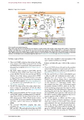
Viral recognition and antiviral responses.
When viruses enter cells they are degraded into short fragment of double-stranded RNA (dsRNA), single-stranded RNA (ssRNA), 5’ triphosphate
ssRNA or CpG DNA. There are three main groups of sensors that detect these fragments: Toll-like receptors (TLRs, see green box), retinoic acid-
inducible gene I (RIG-I)-like receptors (RLRs, see yellow box) and nucleotide oligomerization domain (NOD)-like receptors (NLRs, see pink box).
These different receptors then activate different signalling systems to induce transcription of the inflammatory cytokines and interferon as described
in the text. The information used to construct this figure was taken from McCartney and Colonna (2009) and Takeuchi and Akira (2009).
Toll-like receptors (TLRs) leus where they contribute to the transcription of the
type I interferons (IFN-α and IFN-β).
1. Those viral PAMPs, which are directed into the endo-
some, interact with Toll-like receptors (TLR3, TLR7/8 Retinoic acid-inducible gene I (RIG-I)-like receptors
and TLR9) that are located in the endosomal membrane (RLRs)
(see green box on the left of Module 2: Figure virus re- 6. Those viral PAMPs that are directed into the cytoplasm
cognition). interact with cytoplasmic receptors such as the RIG-I-
2. The TLR9, which responds to CpG DNA, and the like receptors (RLRs) (see yellow box in the middle of
TLR7/8 that bind ssRNA both recruit the adaptor Module 2: Figure virus recognition).
protein MyD88. The latter then acts through TRAF6, 7. The two main RLRs are retinoic acid-inducible gene
which is one of the tumour necrosis factor (TNF)- I (RIG-I) itself and melanoma-associated gene 5
receptor-associated factor (TRAF) family, to stimulate (MDA5). These two receptors have a characteristic
the IKKαβ. The latter then activates the transcription DExD/H box helicase domain that is responsible
factor NF-κB through the Toll receptor signalling path- for binding double-stranded RNA (dsRNA). RIG-I
way described in more detail earlier (Module 2: Figure can also respond to 5’ triphosphate ssRNA. These
Toll receptor signalling). two receptors also have a pair of N-terminal caspase
3. The NF-κB is imported into the nucleus where it func- recruitment domains (CARD), which are the func-
tions to induce the transcription of both the inflam- tional transducing components that signal to down-
matory cytokines and the genes for type I interferon stream elements. The LGP2 (laboratory of genetics and
(IFN). physiology-2), which also has a helicase domain, can
4. When activated by dsRNA, the TLR3 receptor interacts also bind dsRNA, but it lacks the CARD domains and
with the TIR-domain-containing adaptor protein indu- thus fails to transduce cellular signals but may act as a
cing IFN-β (TRIF). The latter then activates the two negative regulator of MDA5 and RIG-I.
IKK-related proteins TBK1 [TRAF-family member- 8. The CARD domains on MDA5 and RIG-I inter-
associated NF-κB activator (TANK)-binding protein] act with the IFN-β promoter stimulator-1 (IPS-1),
and IKKε, which is also known as inducible IKK which also contains an N-terminal CARD domain
(iIKK). and is attached to the outer mitochondrial membrane.
5. The activated TBK1/IKKε complex then phos- IPS1 is also known as MAVS (mitochondrial anti-
phorylates the interferon-regulatory factors (IFRs) viral signalling), VISA (virus-inducing signalling ad-
IRF3 and IRF7. When phosphorylated, these two IRFs aptor) or CARDIF (CARD adaptor inducing IFN-
form homodimers that are then imported into the nuc- β). The IPS1 protein then stimulates the TBK1/IKKε
C 2012 Portland Press Limited www.cellsignallingbiology.org