
Page 93 - 85 cell signalling pathways
P. 93
Cell Signalling Biology Michael J. Berridge Module 2 Cell Signalling Pathways 2 93
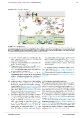
Module 2: Figure Toll receptor signalling
LBP
TLR4
1
CD14 LPS
Src
PLC 1 MyD88 IRAK-1 P P Ubiquitin
4
Ubc13/
2+ 11 TIRAP IRAK-1 Uev1a
Ca P 3 A20
IRAK-4 TRAF6 10
P CYLD
2 TAB1-3 Proteasome
+ TRAF6 TAK1
P NEMO
NFAT P IKK P P
P IKK IB
6 P 8
CaN 5 7
IB P P
p50 p65 IB
JNK p38
p50 p65
NF- B
Pro-apoptotic genes Import Inflammatory cytokines
nur77, Gadd45g, 9 TNF- IL-6; IL-1
Ddit3, Tia
P
JUN Fos p50 p65 Immunoregulators
NFAT
Interleukin-2 TGF- prostaglandins
CRE B PDE4a
The Toll receptor signalling pathway.
The lipopolysaccharide (LPS) that initiates this signalling pathway binds first to an extracellular LPS-binding protein (LBP) and to CD14, which is a
glycosylphosphatidylinositol-anchored membrane glycoprotein, and this complex carries the LPS to the Toll-like receptor 4 (TLR4). The activated TLR4
then recruits a signalling complex to relay information to both the p38 and nuclear factor κB(NF-κB) signalling pathways to induce the transcription
of a number of inflammatory cytokines and immunoregulators as described in the text.
6. The other action of TAK1 is to phosphorylate the such as interleukin-2 and various pro-apoptotic genes
IKKβ component of the inhibitor of NF-κB(IκB) (nur77, Gadd45g, Ddit3 and Tia1)(Module 2: Figure
kinase (IKK) α/IKKβ dimer. Toll receptor signalling). This Ca 2 + pathway has been
7. The phosphorylated IKKβ then acts to phosphorylate described in dendritic cells, but appears to be absent
IκBα to liberate the NF-κB. in macrophages.
8. The phosphorylated IκBα is then susceptible to ubi-
quitination by the Skp1/cullin/F-box (SCF) ubiquitin This signalling pathway is particularly evident on res-
ligase. The polyubiquitinated IκBα is then sent to the ident macrophages (Module 11: Figure macrophage sig-
proteasome, where it is degraded, nalling) and mast cells (Module 11: Figure mast cell sig-
9. The NF-κB enters the nucleus, binds to the κBsite nalling) that respond to invading pathogens. Mutations in
and activates the genes that code for the inflammatory MyD88 have been linked to diffuse large B cell lymphoma
cytokines and immunoregulators that contribute to (DLBCL).
inflammatory responses (Module 11: Figure inflam-
mation).
10. An important aspect of the recovery of this signalling Virus recognition and antiviral responses
cascade after LPS is withdrawn, is the removal of The recognition and initiation of antiviral responses, which
the ubiquitin scaffolds that hold together the trans- is a part of the innate immune system, depend upon the ac-
ducing complex. The deubiquitinating enzymes A20 tivation of a number of signalling pathways such as the Toll
and CYLD are particularly active in removing the ubi- receptor signalling pathway. When viruses enter cells they
quitin chains. This ubiquitin signalling system thus are broken down into fragments such as double-stranded
plays an essential role in the processing of informa- RNA (dsRNA), single-stranded RNA (ssRNA), 5’ tri-
tion by this Toll receptor signalling pathway. phosphate ssRNA or CpG DNA, which are located either
11. In addition to functioning as a coreceptor to transfer in the cytoplasm or within the endosomal compartment
LPS to TLR4, CD14 may also be capable of activat- (Module 2: Figure virus recognition). These fragments are
ing a Ca 2 + signalling pathway. The CD14 activates examples of the pathogen-associated molecular patterns
Src that is coupled to phospholipase Cγ1(PLCγ1), (PAMPs) that are responsible for triggering a variety of in-
which is then capable of triggering an influx of ex- flammatory responses (Module 11: Figure formation and
ternal Ca 2 + through an unknown mechanism. The action of PAMPs). The following sequence of reactions
increase in Ca 2 + stimulates calcineurin (CaN) to de- describes how three main groups of receptors detect these
phosphorylate the transcription factor NFAT that viral PAMPs to initiate a number of antiviral signalling
enters the nucleus to activate expression of genes pathways (Module 2: Figure virus recognition):
C 2012 Portland Press Limited www.cellsignallingbiology.org