
Page 92 - 85 cell signalling pathways
P. 92
Cell Signalling Biology Michael J. Berridge Module 2 Cell Signalling Pathways 2 92
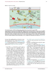
Module 2: Figure NF-κB activation
TNF
TNF TNF
TNFR
1
D D D D D
D D D D D Ubc13/ D D D D Ubc13/ 10
2 Uev1a Ubiquitin 2 2 Uev1a A20 Ubiquitin
F F F
TRADD A 2 A A CYLD
R R R
T ASK1 T T
RIP1 RIP1
TAB1-3
JNK IKK NEMO TAB1-3
p38 IKK TAK1
P
TAK1
5 4 3
P P 6 P P NEMO
Proteasome IB IB IB IKK IKK
p50 p65 p50 p65 p50 p65
7
p50 p65 NF- B 9 IB
Export
Import
IB
8
IL-6
CYLD
p50 p65
A20
B HIF-1
The ‘classical’nuclear factor κB(NF-κB) signalling pathway activated by the tumour necrosis factor receptor (TNFR).
The p50 and p65 isoforms of the nuclear factor κB(NF-κB)/Rel family form the NF-κB dimer that is activated in the tumour necrosis factor α (TNFα)
signalling pathway. The activated TNF receptor (TNFR) recruits a signalling complex to the membrane (Steps 2--4), which contains the inhibitor of
NF-κB(IκB) kinase (IKK) α/IKKβ dimer that is responsible for phosphorylating the IκBα subunit that retains p50/p65 in the cytoplasm (Step 5). When
the IκBα is phosphorylated, it is ubiquitinated and degraded by the proteasome (Steps 6 and 7). The p50/p65 homodimer is imported into the nucleus
(Step 8), where it activates a large number of genes. One of these genes codes for IκBα, which sets up a negative-feedback loop by exporting
p50/p65 from the nucleus (Step 9). Adapted from Trends Biochem. Sci., Vol. 30, Viatour, P., Merville, M.-P., Bours, V. and Chariot, A., Phosphorylation of
NF-κBand IκB proteins: implications in cancer and inflammation, pp. 43--52. Copyright (2004), with permission from Elsevier; see Viatour et al. 2005.
for many other signalling pathways, information is trans- factor (TNF)-receptor-associated factor (TRAF) fam-
ferred through both protein--protein interactions and pro- ily that has a critical role to play in the next series of
tein phosphorylation reactions. The ubiquitin signalling reactions.
system also has an important role in orchestrating this Toll 3. The IRAK-1 and TRAF6 dissociate from the receptor
receptor signalling pathway as shown in the following se- and move in to the cytoplasm.
quence (Module 2: Figure Toll receptor signalling): 4. TheTRAF6is aRINGdomainE3 ubiquitin lig-
ase that associates with the heterotrimeric ubiquitin-
conjugating (E2) complex that contains Ubc13 and
1. The TLR4 is a transmembrane protein that has
Uev1A. This is a K63 ubiquitinating complex that
leucine-rich repeats in its ectodomain, while the
results in the autoubiquitination of TRAF6. The
cytoplasmic domain has a Toll/interleukin 1 (IL-
ubiquitinated TRAF6 then binds the transforming
1) receptor (TIR) domain. The lipopolysaccharide
growth factor β activated kinase-1 (TAK1) and the
(LPS) that initiates this signalling pathway binds first
TAK1-binding (TAB) proteins 1 to 3 (TAB1-3). The
to an extracellular LPS-binding protein (LBP) and
multisubunit cytoplasmic complex containing the in-
to CD14, which is a glycosylphosphatidylinositol-
hibitor of NF-κB(IκB) kinase (IKK) α/IKKβ dimer
anchored membrane glycoprotein, and this complex
and the regulatory NF-κB essential modifier (NEMO)
carries the LPS to the Toll-like receptor 4 (TLR4).
subunit are also drawn into the complex. The result-
2. The TIR domain on the TLR4 receptor forms ho-
ing activation of TAK1 is then responsible for relaying
mophilic interaction with the TIR domain of the
information out to other components of the signalling
adaptor proteins TIR adaptor protein (TIRAP) and
pathways.
MyD88. The latter is specific for certain TLRs, such
as TLR4, but not others. The other end of these ad- 5. The TAK1 activates both the c-Jun N-terminal kinase
aptors have a death domain that draws in the IL- (JNK) pathway and the p38 pathway (Module 2: Fig-
1 receptor-associated kinases 1 and 4 (IRAK-1 and ure MAPK signalling). The JNK and p38 function
IRAK-4), which undergoes an autophosporylation re- to phosphorylate transcription factors such as AP-
action that enables them to bind to another adaptor 1, which binds to the cyclic AMP response element
called TRAF6, which belongs to the tumour necrosis (CRE) site.
C 2012 Portland Press Limited www.cellsignallingbiology.org