
Page 188 - 84 human physiolofy part-1
P. 188
Chapter 10
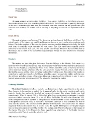
16.Renal papilla
17.Renal column
Renal Vein
The renal veins are veins that drain the kidney. They connect the kidney to the inferior vena cava.
Because the inferior vena cava is on the right half of the body, the left renal vein is generally the longer
of the two. Unlike the right renal vein, the left renal vein often receives the left gonadal vein (left
testicular vein in males, left ovarian vein in females). It frequently receives the left suprarenal vein as
well.
Renal Artery
The renal arteries normally arise off the abdominal aorta and supply the kidneys with blood. The
arterial supply of the kidneys are variable and there may be one or more renal arteries supplying each
kidney. Due to the position of the aorta, the inferior vena cava and the kidneys in the body, the right
renal artery is normally longer than the left renal artery. The right renal artery normally crosses
posteriorly to the inferior vena cava. The renal arteries carry a large portion of the total blood flow to
the kidneys. Up to a third of the total cardiac output can pass through the renal arteries to be filtered by
the kidneys.
Ureters
The ureters are two tubes that drain urine from the kidneys to the bladder. Each ureter is a
muscular tube about 10 inches (25 cm) long. Muscles in the walls of the ureters send the urine in small
spurts into the bladder, (a collapsible sac found on the forward part of the cavity of the bony pelvis that
allows temporary storage of urine). After the urine enters the bladder from the ureters, small folds in
the bladder mucosa act like valves peventing backward flow of the urine. The outlet of the bladder is
controlled by a sphincter muscle. A full bladder stimulates sensory nerves in the bladder wall that relax
the sphincter and allow release of the urine. However, relaxation of the sphincter is also in part a
learned response under voluntary control. The released urine enters the urethra.
Urinary Bladder
The urinary bladder is a hollow, muscular and distendible or elastic organ that sits on the pelvic
floor (superior to the prostate in males). On its anterior border lies the pubic symphysis and, on its
posterior border, the vagina (in females) and rectum (in males). The urinary bladder can hold
approximately 17 to 18 ounces (500 to 530 ml) of urine, however the desire to micturate is usually
experienced when it contains about 150 to 200 ml. When the bladder fills with urine (about half full),
stretch receptors send nerve impulses to the spinal cord, which then sends a reflex nerve impulse back
to the sphincter (muscular valve) at the neck of the bladder, causing it to relax and allow the flow of
urine into the urethra. The Internal urethral sphincter is involuntary. The ureters enter the bladder
diagonally from its dorsolateral floor in an area called the trigone. The trigone is a triangular shaped
area on the postero-inferior wall of the bladder. The urethra exits at the lowest point of the triangle of
the trigone. The urine in the bladder also helps regulate body temperature. If the bladder becomes
completely void of fluid, it causes the patient to chill.
188 | Human Physiology