
Page 187 - 84 human physiolofy part-1
P. 187
The Urinary System
urea which is the primary nitrogenous end product of metabolism in humans. The liver turns the
ammonia into urea because it is much less toxic. We can also excrete some ammonia, creatinine and
uric acid. The creatinine comes from the metabolic breakdown of creatine phospate (a high-energy
phosphate in muscles). Uric acid comes from the break down of necloetides. Uric acid is insoluble and
too much uric acid in the blood will build up and form crystals that can collect in the joints and cause
gout.
Secretion of Hormones The endocrine system has assistance from the kidney's when releasing
hormones. Renin is released by the kidneys. Renin leads to the secretion of aldosterone which is
released from the adrenal cortex. Aldosterone promotes the kidneys to reabsorb the sodium (Na+) ions.
The kidneys also secrete erythropoietin when the blood doesn't have the capacity to carry oxygen.
Erythropoietin stimulates red blood cell production. The Vitamin D from the skin is also activated with
help from the kidneys. Calcium (Ca+) absorption from the digestive tract is promoted by vitamin D.
CC: Chapter Check: Name the role of the kidneys and how they work?
Organs in the Urinary System
Kidneys And Their Structure
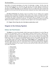
The kidneys are a pair of bean shaped, reddish brown organs about the size of your fist. They are
covered by the renal capsule, which is a tough capsule of fibrous connective tissue. Adhering to the
surface of each kidney is two layers of fat to help cushion them. There is a concaved side of the kidney
that has a depression where a renal artery enters, and a renal vein and a ureter exit the kidney. The
kidneys are located at the rear wall of the abdominal cavity just above the waistline, and are protected
by the ribcage. They are considered retroperitoneal, which means they lie behind the peritoneum. There
are three major regions of the kidney, renal cortex, renal medulla and the renal pelvis. The outer,
granulated layer is the renal cortex. The cortex stretches down in between a radially striated inner layer.
The inner radially striated layer is the renal medulla. This contains pyramid shaped tissue called the
renal pyramids, separated by renal columns. The ureters are continuous with the renal pelvis and is the
very center of the kidney.
1. Renal pyramid
2. Interlobar artery
3. Renal artery
4. Renal vein
5. Renal hylum
6. Renal pelvis
7. Ureter
8. Minor calyx
9. Renal capsule
10.Inferior renal capsule
11.Superior renal capsule
12.Interlobar vein
13.Nephron
14.Minor calyx
15.Major calyx
Wikibooks | 187