
Page 76 - 85 cell signalling pathways
P. 76
Cell Signalling Biology Michael J. Berridge Module 2 Cell Signalling Pathways 2 76
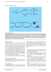
Module 2: Figure GSH/GSSG couple
GSSG
Reduction
GSH Glu Cys Gly
S
Glu Cys Gly S
SH Glu Cys Gly
Oxidation
*
S
H O 2 GSH Grx-S 2
2
SH
S
H O GSSG Grx-(SH) 2 S
2
The GSH/GSSG redox couple.
GSH is a tripeptide consisting of glutamic acid, cysteine and glycine. In its oxidized state, two molecules of GSH are joined together through a
disulphide bond to form GSSG. This is the most abundant redox couple in the cell. The state of this couple can be determined by measuring the
half-cell reduction potential (E -hc ). Under normal reducing conditions, this potential is high, i.e. −240 mV, and this seems to be associated with cell
proliferation. Differentiation seems to occur at lower potentials (−200 mV), whereas still lower potentials of −170 mV favour apoptosis. At these lower
potentials, where there is an alteration in the redox balance, the build-up of GSSG within the cell can reverse the operation of the glutaredoxin system
that functions normally in the recovery of oxidation-sensitive processes. GSSG interacts with reduced glutaredoxin [Grx-(SH) 2 ] to form oxidized Grx-S 2 ,
and this disulphide bond can be transferred to oxidize target proteins.
Glutathione (GSH) peroxide (H 2 O 2 ) to water, thus curtailing its messenger ac-
Glutathione (GSH) is a redox buffer that regulates the tion. PrxI and PrxII are cytosolic, whereas Prx III is found
cellular redox balance (Module 2: Figure GSH/GSSG on mitochondria and PrxIV is on the endoplasmic retic-
couple). GSH is a tripeptide consisting of glutamic acid, ulum. The operation of the catalytic cycle goes through the
cysteine and glycine. It is synthesized by two enzymes. following steps (Module 2: Figure peroxiredoxin catalytic
First, there is glutamate cysteine ligase (GCL), which is cycles):
made up of two subunits: a GCL catalytic subunit (GCLC)
and a GCL modifier subunit (GCLM). The GCLC car-
1. H 2 O 2 is generated near the plasma membrane when
ries out an ATP-dependent condensation reaction between
the PtdIns3,4,5P 3 (PIP 3 ) formed by receptor activation
cysteine and glutamate to form gamma-glutamylcysteine.
Secondly, a GSH synthetase (GSS), which is also known as stimulates NADPH oxidase.
GSH S-transferase (GST), adds a glycine to the dipeptide 2. H 2 O 2 interacts with the reduced cysteine residues
gamma-glutamylcysteine to form GSH. (Cys-SH) in the N-terminal regions of the thioredoxin
A dysregulation of GSH metabolism has been implic- (Trx) dimers to form two oxidized sulphenic residues
ated in schizophrenia (Module 12: Figure schizophrenia). (Cys-SOH).
3. The Cys-SOH can then interact with the conserved
Cys-SH on the C-terminal regions of the neighbouring
Glutathione peroxidase (GPx) dimer to form two intermolecular disulphides.
The glutathione peroxidase (GPx) family uses the reducing 4. The Prx disulphide is converted back into the reduced
power of glutathione to convert H 2 O 2 into water (Module form by Trx, which is regenerated by thioredoxin re-
2: Figure H 2 O 2 metabolism). ductase (Module 2: Figure recovery of protein oxida-
tion).
Peroxiredoxin (Prx) 5. The sulphenic residues formed by Reaction 2 can un-
The peroxiredoxins (PrxI to PrxIV) are a family of small dergo hyperperoxidation by interacting with further
antioxidant proteins that function to metabolize hydrogen molecules of H 2 O 2 to form the sulphinic acid residues.
C 2012 Portland Press Limited www.cellsignallingbiology.org