
Page 72 - 85 cell signalling pathways
P. 72
Cell Signalling Biology Michael J. Berridge Module 2 Cell Signalling Pathways 2 72
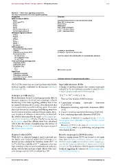
Module 2: Table redox signalling components
The major components of the redox signalling pathway
Component Comment
NADPH oxidases (NOXs)
NOX1 Inducible enzyme found in colon and smooth muscle
NOX2 (gp91 phox ) Major NOX in phagocytes
NOX3 Foetal kidney
NOX4 Widespread
NOX5 Brain, spleen and sperm
DUOX1 (dual oxidase 1) A Ca 2 + -sensitive isoform
DUOX2 (dual oxidase 2) A Ca 2 + -sensitive isoform
NOX/DUOX regulatory factors
p47 phox
p67 phox
p40 phox
p22 phox
NOXO 1
NOXA 1
Rac1/Rac2
ROS metabolism
Superoxide dismutase (SOD)
Catalase Localized in peroxisomes
Glutathione peroxidase (GPx) Localized in cytosol and mitochondria
Peroxiredoxin (Prx)
Prx I--IV (2-Cys) I and II in cytosol; III in mitochondria; IV in endoplasmic reticulum
Prx V (atypical 2-Cys)
Prx VI (1 Cys)
Thiol-containing proteins/peptides
Glutathione (GSH)
Glutaredoxin (Grx)
Thioredoxin (Trx)
Trx-1
Trx-2 Mitochondria-specific
Reductases
Glutathione reductase
Glutaredoxin reductase
Thioredoxin reductase (TrxR)
Sulphiredoxin (Srx) Catalyses reduction of hyperperoxidized proteins
(H 2 O 2 ). This conversion can occur spontaneously, but the Superoxide dismutase (SOD)
reaction is greatly accelerated by the enzyme superoxide A family of metalloproteinases that converts superoxide
dismutase (SOD). radical (O 2 −• ) into hydrogen peroxide (H 2 O 2 )(Module
2: Figure plasma membrane ROS formation):
Hydrogen peroxide (H 2 O 2 ) 2O 2 −• + 2H + → H 2 O 2 + O 2
Much attention is focused on hydrogen peroxide (H 2 O 2 ) There are four families of SOD enzymes:
because it appears to be the primary messenger molecule
functioning in the redox signalling pathway. Since it has • Copper/zinc-containing superoxide dismutases
no unpaired electrons, H 2 O 2 is not a free radical and thus (CuZnSODs)
is not a particularly powerful oxidizing agent. This means • Manganese-containing superoxide dismutases (MnS-
that it can function as a messenger by diffusing away from ODs)
its site of action to interact with more distant targets. How- • Copper-containing superoxide dismutases (CuSODs)
ever, its sphere of influence is restricted by its short half- • Iron-containing superoxide dismutases (FeSODs)
life, which is determined by the rapid reactive oxygen spe-
cies (ROS) metabolism of H 2 O 2 .The H 2 O 2 may also act Expression of MnSOD is regulated by the FOXO3a
as a paracrine signal that diffuses away from stimulated transcription factor (Module 4: Figure FOXO control
cells to alter the activity of neighbouring cells. Release of mechanisms).
H 2 O 2 during wound healing recruits leucocytes as part of Mutation of SOD is the cause of amyotrophic lateral
the inflammatory response. sclerosis (ALS), which is a debilitating and progressive
neurological disease.
•
Hydroxyl radical (OH ) Reactive oxygen species (ROS) formation
While H 2 O 2 is relatively benign, it can be converted into Reactive oxygen species (ROS) are formed at two main
the highly toxic hydroxyl radical (OH ) through a re- sites: there is plasma membrane reactive oxygen species
•
duction process catalysed by transition metals (Fe 3 + or (ROS) formation and mitochondrial reactive oxygen spe-
Cu 2 + ). OH has a half life of 10 − 9 s indicative of its very cies (ROS) formation (Module 2: Figure sites of ROS
•
high reactivity in that it reacts immediately and indiscrim- formation). This production of ROS appears to be highly
inately with the first molecule it finds. Much of the oxid- localized suggesting the existence of reactive oxygen spe-
ative damage cause by ROS is mediated by OH . cies (ROS) microdomains.
•
C 2012 Portland Press Limited www.cellsignallingbiology.org