Page 10 - 68 stomac-duodenum-&-small-intestine59-65_opt
P. 10

Neonatal Intestinal Obstruction  377

                                                                 are present in the submucosal layer of the intestine and migrate from
                                                                 the neural crest distally along the bowel to reach the rectum at about
                                                                 7–10 weeks’ gestation. HD is the congenital absence of neuroganglion
                                                                 cells; consequently, the peristaltic relaxation phase is absent distally,
                                                                 and the affected intestine does not appropriately relax, causing a func-
                                                                 tional obstruction. The extent of the aganglionic segment varies with
                                                                 each patient, but extends from the distal rectum proximally. The level
                                                                 at which the proximal but healthy bowel starts to dilate is called the
                                                                 transition zone (Figure 61.5).
                                                                   The genetic defects responsible for HD consist of abnormalities on
                                                                 more than one chromosome and include the RET proto-oncogene, located
                                                                 at chromosome 10q11.21. RET interacts with a protein termed EDNRB,
                                                                 encoded by the gene EDNRB, which is located on chromosome 13.
                                                                 Anorectal malformation
                                                                 At 4 to 6 weeks’ gestation, the hindgut separates into the urogenital sinus
                                                                 and the anorectum, which then undergoes canalisation. The distal third of
                                                                 the anus develops from ectoderm and becomes the anal pit, whereas the
                                                                 proximal portion of the anal canal is derived from mesoderm. An anal
                                                                 membrane covers the canal until 8 weeks’ gestation, when it perforates
                                                                 and becomes a patent anus. Imperforate anus results if this sequence of
                                                                 events occurs improperly.
                                                                   In summary, conditions of NIO include:
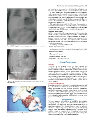
          Figure 61.1: Radiograph showing ground-glass appearance of meconium ileus.
                                                                  • hernia (inguinal, internal);
                                                                  • atresia, stenosis, web (oesophageal, duodenal, jejunoileal, colonic);
                                                                  • anorectal malformation;
                                                                  • Hirschsprung’s disease;
                                                                  • meconium ileus or plug; and
                                                                  • malrotation with midgut volvulus.
                                                                               Clinical Presentation
                                                                 History
                                                                 The  history  of  NIO  is  typical  for  the  level  (high/  low)  and  type
                                                                 (mechanical,  functional)  of  obstruction.  High  intestinal  obstruction
                                                                 presents with early vomiting, whereas low intestinal obstruction pres-
                                                                 ents with abdominal distention and later onset vomiting. Feeding intol-
                                                                 erance  with  bile-stained  vomiting,  absent  meconium,  and  abdominal
                                                                 distention are therefore paradigmatic. With late presentation, the symp-
                                                                 tom presentation might change due to complications and malnutrition.
                                                                   On  antenatal  ultrasound,  polyhydramnios  is  a  common  feature.
                                                                 Intrauterine bowel dilatation may also be noted if scanned after 24
          Figure 61.2: Meconium peritonitis with calcification and pseudocyst due to in   weeks’ gestation.
          utero perforation.
                                                                 Physical Examination
                                                                 Depending  on  the  level  of  intestinal  obstruction,  the  physical  find-
                                                                 ing is that of a distended abdomen. Within the African context, these
                                                                 babies  often  present  late  with  aspiration  pneumonia,  malnutrition,
                                                                 and final events such as intestinal perforation and sepsis. A careful
                                                                 examination and search for typical signs (e.g., abdominal distention,
                                                                 peristaltic waves across the abdomen, absent anus or perianal fistulas,
                                                                 severe distention, and malnutrition in HD) usually reveal the appro-
                                                                 priate suspected diagnosis.
                                                                                   Investigations
                                                                 The  most  important  and  useful  test  in  any  NIO  is  the  abdominal
                                                                 radiograph (AXR). A single supine and lateral shoot-through is usu-
                                                                 ally sufficient. The distribution of the air contrast directs one to the
                                                                 appropriate diagnosis. Very distended loops of bowel are indicative
                                                                 of  atresia.  Long-standing  drainage  from  a  nasogastric  tube  (NGT),
                                                                 however, could make such a diagnosis difficult. A duodenal atresia
                                                                 could in this way be missed purely by the duodenal bulb being col-
          Figure 61.3: Meconium ileus with perforation.          lapsed from the ongoing drainage. A repeat radiograph with injection
                                                                 of some air through the NGT facilitates the diagnosis.
   5   6   7   8   9   10   11   12   13   14   15