
Page 11 - 67 abdominal-wall56-58_opt
P. 11
CHAPTER 58
Inguinal and Femoral Hernias
and Hydroceles
Francis A. Abantanga
Kokila Lakhoo
Introduction
In general, a hernia is defined as a protrusion of a portion of an organ Groin hernias
or tissue through an abnormal opening (defect) in the cavity containing
Direct inguinal Femoral hernia
it. In children, the abnormal defect, which is congenital, is usually at
hernia 0.5–1.0% .05%
the internal inguinal ring.
Indirect inguinal
Groin hernias and hydroceles are extremely common conditions in hernia 99.9%
infancy and childhood and form a large part of the general paediatric
surgical practice. Inguinal hernias (IHs) and hydroceles in infants and
children are overwhelmingly congenital, although a vast majority
are noticed after the neonatal period. Most hydroceles in infants and Inguinal Inguinoscrotal
children do not present any urgent problems.
Demographics
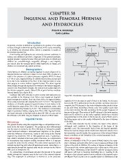
Groin hernias in children are mainly inguinal in nature (Figure 58.1).
Inguinal hernias are indirect in nature in more than 99% of cases as a Reducible Irreducible
result of the presence of a patent processus vaginalis (PPV). In about
0.5–1% of cases, inguinal hernias in children may be direct and are said
to be due to the weakness of the floor of the inguinal canal or occur
after surgery to correct indirect inguinal hernias. The direct inguinal Incarcerated Strangulated
hernia bulges through the inguinal floor medial to the inferior epigastric
vessels in the Hasselbach’s triangle; the indirect hernia arises lateral to
the inferior epigastric vessels. About 0.5% of groin hernias constitute Obstructed
femoral hernias (see Figure 58.1).
Incidence data with reference to groin hernias and hydroceles are Figure 58.1: Classification of groin hernias.
not available in the literature from Africa; most reports are hospital-
based retrospective studies. Such data from Africa on inguinal hernias
show a male-to-female ratio ranging from 2.2:1 to 16.6:1. The reported vaginalis (PV) in the male or canal of Nuck in the female. As the testes
incidence of clinically apparent inguinal hernias in term babies in the descend, the PV is pushed ahead into the scrotum, and when descent is
world literature ranges from 1% to 5% in large paediatric series, with complete, the PV proximal to the testis obliterates either shortly before
males outnumbering females by 3–10:1. The incidence is considerably or just after birth, becoming a fibrous cord. This usually occurs later
higher in premature babies, ranging from 7% to 35%. Inguinal hernias on the right side than the left, accounting for the greater frequency of
are found variously on the right side in about 60–70% of cases and on hernias on the right. The portion of the PV adjacent to the testes remains
the left side in 25–30%. They are bilateral in about 5–10% of cases. patent and is referred to as the tunica vaginalis (which has a visceral
and parietal layer) of the testes. In the female, the canal of Nuck ends in
Inguinal Hernia the labium majus and is also usually obliterated by the time of delivery
Embryology of the baby.
As the testis descends into the scrotum, the layers of the anterior
The gonads develop along the urogenital ridge as retroperitoneal struc-
abdominal wall contribute to the formation of the layers of the
tures by the 6th week of gestation. The gonads are then differentiated
spermatic cord. The transversalis fascia forms the internal spermatic
into the testes or ovaries by the 7th to 8th week of intrauterine growth
fascia; the internal oblique and the transversus abdominis muscles form
under hormonal influence. Retroperitoneal migration of the gonads,
the cremasteric muscle; finally, the aponeurosis of the external oblique
under the influence of hormones, results in their being at the internal
muscle contributes to the formation of the external spermatic fascia.
inguinal ring around the 12th to 14th gestational week. A gubernacu-
lum, which is attached to the lower poles of the testes, is a condensation Pathophysiology
of mesenchyme that contains cordlike structures within it. It appears Failure of obliteration of the PV (or canal of Nuck) leads to the occur-
to guide the testes into the scrotum. The testes remain quiescent at the rence of hernias and hydroceles, the two most common problems of the
internal inguinal ring until about 28 gestational weeks, when there is a region of the groin in children. The variety of degrees of patency of the
rapid descent through the inguinal canal into the scrotum by the 36th to PV account for the various pathologies seen in that region of the groin
40th week of intrauterine life. (Figure 58.2). Obliteration of the distal PV with the proximal portion
An outpouching of peritoneum precedes the descent of the gonad still patent will lead to intestines herniating into it, resulting in the
(testis) through the inguinal canal at the level of the internal inguinal formation of an indirect inguinal hernia confined to the inguinal region
ring. This outgrowth of peritoneum is referred to as the processus (see Figure 58.2C). In the case of complete failure of obliteration of the