
Page 6 - 67 abdominal-wall56-58_opt
P. 6
Disorders of the Umbilicus 353
neutropaenia). The incidence of omphalitis in developing countries performed to rule out any retained omphalomesenteric duct or urachal
is significantly higher (as high as 6%) than in developed countries remnants, which require further work-up. 1,7,13
7
(0.7%). Dermoid Cyst of the Umbilicus
Proper umbilical cord care is important in decreasing the incidence Dermoid cyst of the umbilicus is a rare umbilical mass caused by inclu-
of omphalitis as well as neonatal tetanus (which may or may not be sion of skin epithelium below or within the normal skin of the umbili-
associated with omphalitis). Public health interventions have proven cus. On examination, the umbilicus appears wider and darker in color
effective in decreasing the incidence and death from these infections. In than normal, and shiny. No inflammation is noted unless the cyst is
Nepal, for example, the use of chlorhexidine decreased the incidence of infected. The diagnosis is made at surgery on finding the characteristic
5
omphalitis by 75% and its mortality by 24% compared to dry cord care. toothpaste-like sebaceous material within the umbilical mass. Surgical
More than a half million deaths occur yearly in newborn infants excision is curative.
from neonatal tetanus. A high rate of neonatal tetanus was seen among
the Maasai people in Kenya and Tanzania, who applied cow dung to Omphalomesenteric or Vitelline Remnant
the umbilical stumps of their infants. In one simple health programme During early foetal development, the omphalomesenteric or vitelline
among the Maasai people, the death rate from neonatal tetanus duct serves as a conduit from the yolk sac to the midgut. It normally
decreased from 82 per 1,000 in control groups to 0.75 per 1,000 in completely involutes by the 9th week of foetal life. However, a portion
the intervention group. Part of the success was in finding solutions or all of the duct may fail to involute and present as one of the following:
11
that were culturally applicable and feasible (e.g., if clean water was • An umbilical polyp, as discussed in the next section.
unavailable, they advocated cleaning the stump with milk), obtaining • Meckel’s diverticulum, in which only the diverticulum attached to
support from within the community, and maintaining continued health the ileum has failed to involute. This is the most common vitel-
promotion. line remnant; it most often presents as a lower GI bleed caused by
Patients with omphalitis present with erythema, oedema, and/ ectopic gastric mucosa, but rarely may present as diverticulitis, or it
or purulent drainage from the umbilical stump. Patients may also may function as the lead point for an intussusception.
have systemic signs of sepsis, including lethargy, irritability, poor
feeding, and fever or hypothermia. More extensive disease is seen • A persistent congenital band, which can act as a fixed point around
with necrotising fasciitis or myonecrosis and may also include a which an intestinal volvulus may occur.
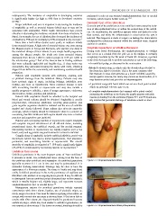
rapidly progressive cellulitis, a peau d’orange appearance, violaceous • A complete omphalomesenteric duct remnant with a patent conduit
discoloration, bullae, crepitus, and petechiae. connecting the umbilicus to the ileum; this usually presents with pink
Patients with omphalitis should be admitted to the hospital and mucosa protruding from the umbilicus (Figures 57.1 and 57.2) and usu-
blood and wound cultures should be obtained. Omphalitis is usually ally minimal but persistent discharge of intestinal contents or stool.
polymicrobial; intravenous antibiotics covering gram-positive and
gram-negative organisms should be initiated and the area of cellulitis
marked and closely followed. Some authors also advocate anaerobic
coverage, which certainly should be instituted if there is a concern of
necrotising fasciitis. Newborns with sepsis should also have a lumbar
puncture and supportive care instituted.
Patients with necrotising fasciitis or myonecrosis require emergent
and complete surgical debridement of all affected tissue, including
preperitoneal tissue, the umbilical vessels, and the urachal remnant.
Necrotising fasciitis or myonecrosis can rapidly progress over a few
hours; early and aggressive surgical treatment is critical to survival.
Complications of omphalitis include umbilical phlebitis, portal vein
thrombosis (which may lead to portal hypertension), liver abscesses,
peritonitis, and necrotising fasciitis or myonecrosis. The overall
mortality of omphalitis is estimated at 7–15% and is significantly higher
(37–87%) if complicated by necrotising fasciitis or myonecrosis. 12
Umbilical Granuloma
Umbilical granuloma is the most frequent cause of “wet umbilicus.”
It presents as moist, raw, reddish-pink tissue arising from the base of
the umbilicus after umbilical cord separation. An umbilical granuloma Figure 57.1: Omphalomesenteric fistula.
typically measures 0.1–1 cm in size and may be pedunculated. It is
nontender (lacking innervation). Drainage may be clear or have the
appearance of a fibrinous exudate. The tissue is friable and may bleed
easily. Umbilical granuloma is due to the persistence of capillary and
fibroblast cells, markers of an ongoing tissue growth. It may be difficult
to distinguish from an umbilical polyp (discussed later in this chapter),
which is usually brighter red, slightly larger, and represents remnant
omphalomesenteric duct or urachal tissue.
7,8
Management options for umbilical granuloma include repeated
cauterisation with silver nitrate, ligation, use of alcoholic wipes, or,
rarely, surgical excision. Care must be taken in applying silver nitrate, as
contact with normal skin can cause a chemical burn. If the lesion fails to
resolve with silver nitrate, the diagnosis should be questioned because
umbilical polyps, which may look similar to umbilical granulomas, do
not respond to silver nitrate. If the lesion is excised, histology should be Figure 57.2: Omphalomesenteric fistula (intraoperative).