
Page 12 - 61 surgical-infection&infestations20-24_opt
P. 12
CHAPTER 22
Haematogenous Osteomyelitis
and Septic Arthritis
Donald E. Meier
Bankole S. Rouma
Haematogenous Osteomyelitis
Haematogenous osteomyelitis (HO) is a common and devastating prob-
1
lem for children in less developed areas of the world due to its frequent
association with sickle cell disease (23–44%), delayed presentation,
misdiagnosis, and undertreatment. Sixty to eighty percent of children
1–5
do not initially present until they have reached the stage of chronic
osteomyelitis. In more medically advanced areas, the spectrum of HO
1–4
has changed significantly in the past few decades with decreased preva-
lence, earlier presentation, better nourished children, increased aware-
ness of HO, improved diagnostic modalities for confirmation, precise
laboratory techniques for microbial identification, and advanced anti-
microbial agents for successful eradication of the infection. In locations
without advanced technology (LWATs), however, little has changed in
the past half century in the presentation or management of children with
HO. Most children in advanced areas undergo successful nonopera-
tive eradication of HO, many with nothing more invasive than a blood
culture or bone aspiration. Most current Western literature concerning
6–8
HO—which concentrates on whether diagnostic aspiration of the bone
marrow is really necessary and whether one powerful antibiotic is bet-
ter than another in the eradication of HO—therefore has little relevance
to practitioners in LWATs who are fortunate if they have materials for
a gram stain and enough antibiotics for a week of oral treatment before
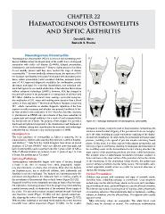
the family runs out of funds. The purpose of this chapter is to provide a Figure 22.1: Pathologic development of haematogenous osteomyelitis.
functional and practical approach to the classification and treatment of
HO in children, taking into consideration the economic and technologic increases in volume, it takes the path of least resistance. Sometimes, this
restraints that are inherent in any medical practice in LWATs.
involves circumferential stripping of the periosteum from one metaphy-
Demographics sis to the other, resulting in a giant sequestrum consisting of the diaphy-
The exact incidence of osteomyelitis in Africa is unknown, but in sis and both metaphyses. At other times, the periosteum perforates under
reported series, children with osteomyelitis represent 7–20% of hospital- pressure, resulting in the spread of pus into muscles and along fascial
ised children. 1–-3 Data from the United Kingdom have shown an annual planes. At this point, it is often confused with primary pyomyositis, and
incidence of 2.9 per 100,000, with boys affected more than girls, and the bony origin is overlooked, resulting in inadequate decompression of
9
half of osteomyelitis cases occurring in the first 5 years of life. In Africa, the medullary canal. As the devascularised cortex is being absorbed, the
1
56% of affected children are between 8 and 11 years of age. In Cote inner surface of the periosteum produces new bone, called involucrum.
d’Ivoire, the median age is 7.2 years. 2 The multiple areas of subperiosteal involucrum formation coalesce to
Aetiology/Pathology form new bone on the inner surface of the periosteum but on the outside
Haematogenous osteomyelitis begins with entry of bacteria through of the sequestrum. If the sequestrum totally resorbs, the patient may
a break in the skin or mucosa from otitis, pharyngitis, respira- recover without a problem, but this rarely occurs. The nonviable, unre-
tory tract infections, or urinary tract infections. Most often the bacteria sorbed sequestrum usually serves as a nidus for recurrent abscesses or
are Staphylococcus, but in sickle-cell children, both Salmonella and chronically draining sinuses.
Staphylococcus are implicated. The bacteria are haematogenously dis- Clinical Presentation
seminated and deposited in the trabecular bone or marrow, usually in Children may present with symptoms and signs of systemic sepsis,
the metaphysis of the proximal tibia or distal femur (Figure 22.1(A)). including fever, irritability, lethargy, or convulsions. Local symptoms
Sluggish blood flow in the metaphysis provides an ideal milieu for bacte- include pain over the affected bone, which can be acute and overwhelm-
rial replication. Increasing pressure from the progressive, intramedullary ing or insidious in onset, and unwillingness to use the affected extremity.
purulent process results in destruction of the endosteal blood supply to The history may include a recent episode of impetigo, otitis, pharyngitis,
the cortex. The pus under pressure escapes outward through Volkmann or respiratory infection. Characteristically, there is tenderness, swelling,
and Haversian canals (Figure 22.1(B)) and then spreads subperioste- redness, or shininess over the affected bone. The adjacent joint usually
ally, stripping the cortex of its periosteal blood supply (Figure 22.1(C)). appears normal.
Without either endosteal or periosteal blood supply, the cortex becomes Most laboratory values are nonspecific. The white blood count
nonviable bone called sequestrum (Figure 22.1(D)). As the exudate (WBC) may be elevated or normal. The erythrocyte sedimentation