
Page 21 - Noninvasive Diagnostic Techniques for the Detection of Skin Cancers
P. 21
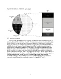
Figure 3. Distribution of all abstracts by study type


               RCT = randomized, controlled trial

                   Over half of the abstracts addressed melanoma detection and diagnosis (60 percent) and 13
               percent addressed BCC or SCC (see Figure 4). The remainder covered skin cancer combinations
               or did not specify the type of skin cancer lesion (see Appendix D, Table D2). In considering
                                                                                                      16
               devices in current use, we reviewed the classification system presented by Marghoob 2003  in
               which the devices are compared by skin imaging depth. This classification system helped to
               clarify the type of information gleaned from the imaging device as well as alternative devices
               designed to capture similar information. For example, photography is considered a quaternary
               device, providing information at the superficial level, while dermoscopy helps to characterize
               lesions at the tertiary level (e.g., cellular aggregates or blood vessels). Neither of these devices is
               designed to delineate specific cellular and subcellular structures. Investigational devices, such as
               confocal microscopy may provide that level of resolution. In general, issues of access,
               availability, and degree of required training, increase with tissue depth. Outcome measures
               reported in the published primary studies are presented in Appendix D, Table D4.


                                                             12