
Page 123 - 85 cell signalling pathways
P. 123
Cell Signalling Biology Michael J. Berridge Module 2 Cell Signalling Pathways 2 123
Module 2: Figure AMPK structure
GBD BD
I C
Enzyme activation by
AMP and/or protein phosphorylation
by an AMPK kinase (LKB1)
GBD BD
2+
Ca + CaMKK + I
P
+ AMP
LKB1 C
Protein +
Protein- P AMP
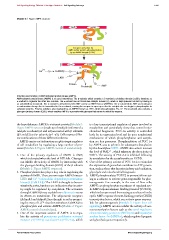
Structure and function of AMP-activated protein kinase (AMPK).
AMP-activated protein kinase (AMPK) is an αβγ heterotrimer. The β-subunit, which contains a C-terminal αγ-binding domain (αγBD), functions as
a scaffold to organize the other two subunits. The α-subunit has an N-terminal catalytic domain (C), which is kept quiescent at rest by binding to
an autoinhibitory domain (I). This α-subunit is activated by both AMP and by an AMPK kinase (AMPKK). The γ-subunit binds AMP and undergoes
a conformation change that is transmitted to the α-subunit, causing the enzyme to open up so that its catalytic site can begin to phosphorylate its
substrate proteins. Enzyme activity is also regulated by an AMPKK known as LKB1, which phosphorylates Thr-172. The β-subunit also contains a
glycogen-binding domain (GBD), which enables AMPK to associate with glycogen that serves to inhibit the enzyme.
the hypothalamus. AMPK is a trimeric protein (Module 2: is a key transcriptional regulator of genes involved in
Figure AMPK structure) made up of multiple isoforms of a metabolism and particularly those that control mito-
catalytic α subunit (α1and α2) associated with β-subunits chondrial biogenesis. PGC-1α activity is controlled
(β1and β2) and γ-subunits (γ1--γ3). Cells express differ- both by its expression level and by post-translational
ent combinations of these different isoforms. mechanisms of which phosphorylation and acetyla-
AMPK carries out its function as a pleiotropic regulator tion are key processes. Phosphorylation of PGC-1α
of cell metabolism by regulating a large number of pro- by AMPK acts to prime it for subsequent deacylation
cesses (Module 2: Figure AMPK control of metabolism): by the deacetylase SIRT1. AMPK also acts to increase
the level of NAD , which enhances the the activity of
+
1. One of the primary regulators of AMPK is AMP, SIRT1. The activity of PGC-1α is inhibited following
which is elevated when the level of ATP falls. Glycogen its acetylation by the acetyltransferase GCN5.
can inhibit the activity of AMPK by interacting with 4. One of the primary actions of PGC-1α is to stimulate
the glycogen-binding domain (GBD) of the β subunit the expression of genes that contribute to ATP genera-
(Module 2: Figure AMPK structure). tion, such as those that function in fatty acid oxidation,
2. Phosphorylation also plays a key role in regulating the glycolysis and mitochondrial biogenesis.
activity of AMPK. There are two major AMPK kinases- 5. AMPK phosphorylates TORC2 to prevent it from act-
LKB1 and Ca 2 + /calmodulin-dependent protein kinase ing as a cofactor to activate genes responsible for gluc-
kinase β (CaMKKβ). The LKB1 is thought to be con- oneogenesis. For example, in the case of liver cells,
stitutively active, but there are indications that its activ- AMPK can phosphorylate transducer of regulated cyc-
ity might be regulated by acetylation. The activation lic AMP response element-binding protein 2 (TORC2),
through CaMKKβ may explain how adiponectin exerts which is then prevented from acting as a cofactor for the
its effects on metabolism. The adiponectin receptors cyclic AMP response element-binding protein (CREB)
(AdipoR1 and AdipoR2) are thought to act by promot- transcription factor, which can activate genes respons-
ing the entry of Ca 2 + that then stimulates CaMKKβ to ible for gluconeogenesis (Module 7: Figure liver cell
phosphorylate and activate AMPK (Module 2: Figure signalling). AMPK can also inhibit the activity of vari-
AMPK control of metabolism). ous transcription factors such as the sterol regulatory
3. One of the important actions of AMPK is to regu- element-binding protein 1c (SREBp1c) and hepatocyte
late the activity of peroxisome-proliferator-activated nuclear factor 4α (HNF4α), which regulate lipogenic
receptor γ (PPARγ) coactivator-1α (PGC-1α),which and glycolytic genes respectively.
C 2012 Portland Press Limited www.cellsignallingbiology.org