
Page 120 - 85 cell signalling pathways
P. 120
Cell Signalling Biology Michael J. Berridge Module 2 Cell Signalling Pathways 2 120
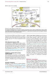
Module 2: Figure ER stress signalling
P e1F-2 Protein synthesis OFF
P P Endoplasmic reticulum
PERK e1F-2 Protein synthesis ON
Protein synthesis
Protein folding
1
Dysfunctional Normal
chaperones chaperones
STRESS
Export
Oligomerization Misfolded 4
protein
2 Excess
3 6
protein
Caspase-12 7
IRE1 P P
APOPTOSIS NF-kB INFLAMMATORY
ATF-6 RESPONSES
Transcription
factors
5
PDI: GRP78:
GRP94; CHOP Interleukins
Calreticulin etc Cytokines
Endoplasmic reticulum (ER) stress signalling pathways.
An accumulation of misfolded proteins or an excessive accumulation of normal proteins activate a number of signalling pathways. Chaperones within
the endoplasmic reticulum (ER) lumen are responsible for folding newly synthesized proteins into their tertiary structures prior to their export to the
Golgi. A variety of stress factors, including a decline in the luminal level of Ca 2 + , results in dysfunctional chaperones and an accumulation of misfolded
proteins that can activate a number of signalling pathways.
nature of the stress. The fact that the ER can up-regulate C/EBP (CCAAT/enhancer-binding protein)-homologous
chaperone levels results in the phenomenon of tolerance, protein 10 (CHOP). Another mechanism depends upon
whereby treatment of cells with low levels of stress stimuli the ER directly activating a subset of caspases during
can make cells much more tolerant to subsequent stressful ER stress. A critical component is caspase 12, which is
stimuli. associated with the ER membrane and is released by
proteolytic cleavage following ER stress. Several mech-
Unfolded protein response (UPR) anisms have been proposed for this activation process.
An accumulation of misfolded proteins induces an unfol- One suggestion is that the stress sensor molecule IRE1
ded protein response (UPR), which switches off ongoing recruits tumour-necrosis-factor-receptor-associated factor
protein synthesis and also activates various transcriptional (TRAF) which then binds to caspase 12, making it sens-
cascades that result in the up-regulation of many of the itive to the Ca 2 + -responsive cysteine protease m-calpain.
key chaperones in an attempt to improve the defective Another suggestion is that the hydrolysis of caspase 12 is
protein packaging machinery (Module 2: Figure ER stress mediated by caspase 7, which is recruited to the membrane
signalling). Activating transcription factor 6 (ATF6) is one during ER stress. An interesting aspect of this mechanism
of the transcription factors activated by the UPR. is that glucose-regulated protein 78 kDa (GRP78) appears
to inhibit this activation process by forming a complex
with caspase 7 and caspase 12. Once caspase 12 is released
Endoplasmic reticulum (ER) overload into the cytosol, it activates a specific cascade involving
response (EOR)
An excessive build-up of proteins, as occurs during viral in- caspase 9 and caspase 3 in a cytochrome c-independent
fections, switches on an endoplasmic reticulum (ER) over- manner.
load response (EOR) that acts through the nuclear factor
κB(NF-κB) signalling cascade to stimulate the release of
interferons and cytokines as part of an inflammatory re- Metabolic messengers
sponse.
There are a number of cellular metabolites that function as
metabolic messengers to integrate the activities of cellular
Activation of apoptosis metabolism and cell signalling (Module 2: Figure meta-
Endoplasmic reticulum (ER) stress signalling pathways bolic messengers). In this context, a metabolic messenger
can also contribute to apoptosis (Module 11: Figure ap- is defined fairly widely: it includes components that are
optosis). For example, one of the UPR pathways depends either a part of, or are derived from, cellular metabolism.
upon the release of the transcription factor activating Metabolism is regulated at many different levels. The most
transcription factor 6 (ATF6), which acts to switch on direct control depends upon feedback processes where
C 2012 Portland Press Limited www.cellsignallingbiology.org