
Page 134 - LECTURE NOTES
P. 134
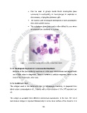
•  Can be seen in groups inside blood monocycles (less

                                            commonly in neutrophils), in macrophages in aspirates or
                                            skin smears, or lying free between cells

                                         •  The nucleus and rod-shaped kinetoplast in each amastigotes

                                            stain dark reddish mauve
                                         •  The cytoplasm stains pale and is often difficult to see when

                                            amastigotes are clustered  in a group.


                                     Figure:  5   Leishmania amastigotes in Giemsa stained skin smear


               5.7.3. Serological diagnosis of cutaneous leshmaniasis

                   Because of the poor antibody response in continuous leishmaniasis serological tests
                   are of little value in diagnosis. There is however a cellular response, which is the

                   basis of the leishmanin, skin tests.


               5.7.4. Leishmanin test

               The antigen used in the leishmanin test (or Montenegro reaction), is prepared from
                                                                                             6
               killed culture promastigotes of L. tropica, with a concentration of 10 x 10  parasites per
               ml.


               The antigen is available from different commercial manufacture. In the test, 10.1 ml of

               well-shaken antigen is injected intradermally in to the inner surface of the forearm. It is


                                                            128