
Page 126 - 84 human physiolofy part-1
P. 126
Chapter 7
WBCs are classified by phenotype which can be identified by looking at the WBCs under a
microscope. The Granular phenotype are able to stain blue. The Agranular phenotype are able to stain
red. Neutrophils make up 50-70% of Granular cells Eosinophils make up 2-4%, and Basophils 0-1%.
Monocytes make up 2-8% of Agranular cells. B and T Lymphocytes make up 20-30%. As you can see,
there is a great deal of differentiation between WBCs. These special cells help our bodies defend
themselves against pathogens. Not only do they help our immune system but they remove toxins,
wastes, and abnormal or damaged cells. Thus, we can say that WBCs' main function is being
Phagocytic which means to engulf or swallow cells.
Platelets
Platelets, also called thrombocytes, are membrane-bound cell
fragments. Platelets have no nucleus, are between one to two
micrometers in diameter, and are about 1/10th to 1/20th as abundant
as white blood cells. Less than 1% of whole blood consists of
platelets. They result from fragmentation of large cells called
Megakaryocytes - which are cells derived from stem cells in the bone
marrow. Platelets are produced at a rate of 200 billion per day. Their
production is regulated by the hormone called Thrombopoietin. The
circulating life of a platelet is 8–10 days. The sticky surface of the
platelets allow them to accumulate at the site of broken blood vessels
to form a clot. This aids in the process of hemostasis ("blood
stopping"). Platelets secrete factors that increase local platelet
aggregation (e.g., Thromboxane A), enhance vasoconstriction (e.g.,
Serotonin), and promote blood coagulation (e.g., Thromboplastin).
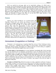
A 250 ml bag of newly collected
platelets.
Hemostasis (Coagulation or Clotting)
Hemostasis is the natural process of stopping blood flow or loss of blood following an injury.
(hemo = blood; stasis = standing). It has three stages: (1) vascular spasm, vasoconstriction, or intense
contraction of blood vessels, (2) formation of a platelet plug and (3) blood clotting or coagulation.
Once the flow of blood has been stopped, tissue repair can begin.
Vascular spasm or Vasoconsriction: In a normal individual, immediately after a blood vessel has
been cut and endothelial cells are damaged, vasoconstriction occurs, thus slowing blood flow to the
area. Smooth muscle in the vessel wall goes through spasms or intense contractions that constrict the
vessel. If the vessels are small, spasms compress the inner walls together and may be able to stop the
bleeding completely. If the vessels are medium to large-sized, the spasms slow down immediate
outflow of blood, lessening the damage but still preparing the vessel for the later steps of hemostasis.
These vascular spasms usually last for about 30 minutes, long enough for the next two stages of
hemostasis to take place.
Formation of a Platelet Plug: Within 20 seconds of an injury, coagulation is initiated. Contrary to
popular belief, clotting of a cut on the skin is not initiated by air or drying out, but by platelets adhering
to and activated by collagen in the blood vessels endothelium. The activated platelets then release the
contents of their granules, which contain a variety of substances that stimulate further platelet
activation and enhance the hemostatic process.
126 | Human Physiology