
Page 242 - AIDSBK23C
P. 242
Page 242
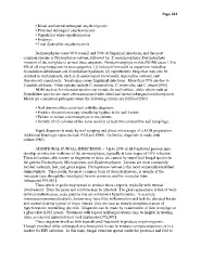
• Distal and lateral subungual onychomycosis
• Proximal subungual onychomycosis
• Superficial white onychomycosis
• Endonyx
• Total dystrophic onychomycosis
Dermatophytes cause 90% toenail and 50% of fingernail infections, and the most
common species is Trichophyton rubrum, followed by T. mentagrophytes. Dermatophyte
invasion of the nail plate is termed tinea unguium. Nondermatophyte molds (NDM) cause 1.5 to
6% of all onychomycosis in two categories: (1) isolated from nails as organisms including
Scytalidium dimidiatum and Scytalidium hyalinum; (2) opportunistic fungi that may also be
isolated as contaminants, such as Scopulariopsis brevicaulis, Aspergillus sydowii, and
Onychocola canadensis. Yeasts may cause fingernail infections. More than 70% are due to
Candida albicans. Other species include C. parapsilosis, C. tropicalis, and C. krusei.[986]
NDM such as Acremonium species can invade the nail surface, while others such as
Scytalidium species are more often associated with distal and lateral subungual onychomycosis.
Molds are considered pathogens when the following criteria are fulfilled:[986]
• Nail abnormalities consistent with the diagnosis.
• Positive direct microscopy visualizing hyphae in the nail keratin.
• Failure to isolate a dermatophyte in the culture
• Growth of >5 colonies of the same mold in at least two consecutive nail samplings.
Rapid diagnosis is made by nail scraping and direct microscopy of a KOH preparation.
Additional histologic stains include PAS and GMS. Definitive diagnosis is made with
culture.[986]
ADDITIONAL FUNGAL INFECTIONS.-- Up to 20% of HIV-infected persons may
develop an infection with one of the dermatophytes, typically at later stages of HIV infection.
These infections, also known as ringworm or tinea, are caused by superficial fungal species in
the genera Trichophyton, Microsporum, and Epidermophyton. Lesions are most commonly
located on hands, feet, and groin region. Trichophyton rubrum is the most commonly identified
dermatophyte. Tinea pedis, the most common form of dermatophytosis, is usually of the
moccasin type, though the interdigital form is common, and the vesicular form
infrequent.[979,987]
Tinea cruris or pedis may spread to produce tinea corporis, typically with truncal
involvement. Even the penis and scrotum may be affected. The gross appearance is that of well-
defined erythematous, scaly patches that are sometimes hyperkeratotic. In severely
immunocompromised patients, lesions may have little inflammation and lack the elevated border
and central clearing typical of tinea; instead, they are seen as sharply marginated areas of
hyperkeratosis resembling dry skin. Diagnosis can be made by examination of skin scrapings on
a glass slide KOH mount. Treatment with topical antifungal creams may be helpful in treating
tinea, as can oral griseofulvin or oral imidazoles.[972,987]
Tinea unguium involves both toenails and fingernails to produce onychomycosis.
Though proximal white subungual onychomycosis is rare in immune competent persons, it is a
marker for HIV infection. In this form, fungal elements spread under the proximal nail fold to