
Page 3 - 68 stomac-duodenum-&-small-intestine59-65_opt
P. 3
370 Infantile Hypertrophic Pyloric Stenosis
the wound and elevated to lift the transverse colon. This manoeuvre Postoperative Management
enables the surgeon to identify the antrum of the stomach. The lower Postoperative nasogastric decompression is not necessary unless the
third of the stomach is then gently elevated using moist gauze to mucosa has been entered and repaired. Several feeding schedules have
deliver the pyloric mass into the wound (Figure 59.3). been advocated after surgery. Traditional structured feeding regimens
2. A vertical incision is then made into the mid anterior surface through as opposed to more rapid initiation and advancing feeding schedules are
the serosa and superficial muscularis, beginning about 1–2 mm from probably unnecessary. Feedings are begun 4 to 6 hours after operation,
the pyloroduodenal junction to a point 0.5 cm into the lower antrum. normally with low-volume balanced electrolyte or dextrose solution
initially, rapidly advanced to full feeds of formula over the next 12- to
3. The underlying firm fibres are then divided using blunt dissection
24-hour period. If the patient vomits, which is common after this pro-
with a clamp, rounded end of a scalpel blade, or special Benson’s
cedure, the same volume feed that caused the emesis can be repeated.
pyloromyotomy spreader. Special care is taken to prevent mucosal
The patient is usually discharged the day after operation.
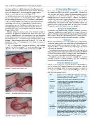
perforation, especially at the lower end of the incision. Upward
protrusion of the gastric mucosa indicates relief of the obstruction Surgical Complications
(Figures 59.4 and 59.5). Intraoperative risks include bleeding, infection, and mucosal perforation.
Mucosal perforation usually occurs at the duodenal end and is Postoperative complications include wound infection and dehiscence in
indicated by the appearance of bilious fluid. When this occurs, repair about 1%. Persistent vomiting beyond 48 hours, thought to be due to gastric
is done by using interrupted fine monofilament long-term absorbable atony, occurs in about 3%. Unrecognized perforation during pyloromy-
sutures placed transversely and covered with omentum. If the closure of otomy is a serious but rare problem demanding immediate reoperation.
the mucosal perforation compromises the pyloromyotomy, which rarely
happens, a fresh pyloromyotomy is done at about 45°–90° of the first Outcome
incision. Air is then instilled through the NGT to check the integrity of The majority of infants go on to make a full recovery postoperatively
the duodenal mucosa. and need no further medical input. After a surgical pyloromyotomy, the
Use of a laparoscopic approach is increasing, with evidence pyloric muscle subsides to a normal size and, when viewed during sub-
supporting its benefits emerging. 18,19 A recent study has shown a safe sequent operations, is usually visible only as a fine line over the pylorus
alternative with a decreased time to full feeds postoperatively. 20 at the site of the myotomy.
Incomplete pyloromyotomy may occur, but it is difficult to diagnose in the
early postoperative phase. Imaging studies done postoperatively are difficult
to interpret and usually not helpful. If complete gastric-outlet obstruction is
present on a contrast study, repeated pyloromyotomy is necessary.
Mortality is rare, but when it occurs, it is usually from fluid and
electrolyte depletion in infants presenting late, and inadequately
corrected electrolyte problems before surgery.
Evidence-Based Research
Evidence on the management of pyloric stenosis in African children is
rare, so clinicians have to depend on literature from the West, where the
Figure 59.3: Operative view of pyloric mass. disease is more frequent. Table 59.1 presents the results of a survey on
the management of IHPS conducted in the United Kingdom and Ireland.
Table 59.1: Evidence-based research.
Title Surgical practice for infantile hypertrophic pyloric stenosis in
the United Kingdom and Ireland—a survey of members of the
British Association of Paediatric Surgeons
Authors Mullassery D, Perry D, Goyal A, Jesudason EC, Losty PD
Institution Academic Department of Pediatric Surgery, The Royal
Liverpool Children’s Hospital (Alder Hey), University of
Liverpool, United Kingdom
Reference J Pediatr Surg 2008; 43:1227–1229
Problem Current practice amongst paediatric surgeons on the
management of infantile hypertrophic pyloric stenosis.
Outcome/ More than half of the surgeons surveyed used umbilical
Figure 59.4: Spreading of the divided pyloric muscle. effect incision for pyloromyotomy, whereas only 15% do the
pyloromyotomy laparoscopically. Fewer than 10% of surgeons
surveyed use the classical right upper quadrant incision for
pyloromyotomy. The study also showed that about half of the
surgeons do not use antiobiotics; however, 70% of those using
the umbilical incision use antibiotics. The study concluded that
umbilical incision and laparoscopic incisions are benchmarks
for surgeons caring for children with infantile hypertrophic
pyloric stenosis.
Historical Acknowledging that IHPS may not be a major workload for
significance/ the paediatric surgeon practicing in Africa, patients with this
comments condition do come in occasionally, especially in major centres,
so paediatric surgeons need to be aware of the current
practices amongst paediatric surgeons who care frequently for
these patients; hence, the importance of this article. Although
there are a lot of variations in the practice, pyloromyotomy
through whatever route remains the gold standard for caring
Figure 59.5: Myotomy with mucosal bulge. for these group of patients.