
Page 28 - 66 thorax49-55_opt
P. 28
Chest Wall Deformities 337
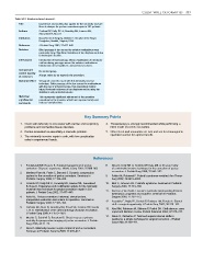
Table 53.2: Evidence-based research.
Title Experience and modification update for the minimally invasive
Nuss technique for pectus excavatum repair in 303 patients
Authors Croitoru DP, Kelly RE Jr, Goretsky MJ, Lawson ML,
Swoveland B, Nuss D
Institution Department of Surgery, Children’s Hospital of the King’s
Daughters, Norfolk, Virginia, USA
Reference J Pediatr Surg 2002; 37:437–445
Problem Blind passage of bar across the anterior mediastinum was
previously done. Significant incidence of bar displacement due
to inadequate fixation.
Intervention Introduction of thoracoscopy allows visualisation of introducer
and bar during passage across the anterior mediastinum.
Introduction of bar stabilisers and pericostal sutures.
Comparison/ No control group.
control (quality
of evidence) A large series by an expert in the procedure.
Outcome/effect Very good cosmetic repair with this minimally invasive
technique. Safer passage of the bar across the mediastinum
with the use of a thoracoscope, thus preventing cardiac
injury. Reduced incidence of bar displacement by using bar
stabilisers and pericostal sutures.
Historical This represents significant refinement of the operative
significance/ procedure by the inventor, which has improved safety and
comments reduced complications.
Key Summary Points
1. Chest wall deformity is associated with cardiac and respiratory 4. Thoracoscopy is strongly recommended while performing a
problems and connective tissue disorders. Nuss repair of pectus excavatum.
2. Pectus excavatum is essentially a cosmetic problem. 5. Other chest wall anomalies are rare and are best managed in
3. The minimally invasive repair is safe, with low complication specialist centres for optimal results.
rates in experienced hands.
References
1. Fonkalsrud EW, Beanes S. Surgical management of pectus 8. Nuss D, Kelly RE Jr, Croitoru DP, Katz ME. A 10-year review
carinatum: 30 years’ experience. World J Surg 2001; 25:898–903. of a minimally invasive technique for the correction of pectus
excavatum. J Pediatr Surg 1998; 33:545–552.
2. Martinez-Ferro M, Fraire C, Bernard S. Dynamic compression
system for the correction of pectus carinatum. Seminars in 9. Folkin AA, Robicsek F. Poland’s syndrome revisited. Ann Thorac
Pediatric Surgery 2008; 17:194–200. Surg 2002; 74:2218–2225.
3. Croitoru DP, Kelly RE Jr, Goretsky MJ, Lawson ML, Swoveland 10. Moir C, Johnson CH. Poland’s syndrome. Seminars in Pediatric
B, Nuss D. Experience and modification update for the minimally Surgery 2008; 17:161–166.
invasive Nuss technique for pectus excavatum repair in 303 11. Duncan J, Van Aalst J. Jeune’s syndrome (asphyxiating thoracic
patients. J Pediatr Surg 2002; 37:437–445.
dystrophy): congenital and acquired. Seminars in Pediatric
4. Kelly RE. Pectus excavatum: historical, clinical picture, Surgery 2008; 17:167–172.
preoperative evaluation and criteria for operation. Seminars in
Pediatric Surgery 2008; 17:181–193. 12. Acastello E, Majluf R, Garrido P, Barbosa LM, Peredo A. Sternal
cleft: a surgical opportunity. J Pediatr Surg 2003; 38:178–183.
5. Cartoski MJ, Nuss D, Goretsky MJ, Proud VK, Croitoru DP, Gustin 13. Abel RM, Robinson M, Gibbons P, Parikh DH. Cleft sternum: case
T, et al. Classification of the dysmorphology of pectus excavatum. report and literature review. Pediatr Pulmonol 2004; 37:375–377.
J Pediatr Surg 2006; 41:1573–1581.
14. Daum R, Zachariou Z. Total and superior sternal clefts in
6. Mueller C, Saint-Vil D, Bouchard S. Chest x-ray as a primary
modality for preoperative imaging of pectus excavatum. J Pediatr newborns: a simple technique for surgical correction. J Pediatr
Surg 2008; 43:71–73. Surg 1999; 34:408–411.
7. Nuss D. Minimally invasive surgical repair of pectus excavatum.
Seminars in Pediatric Surgery 2008; 17:209–217.