
Page 6 - 63 craniocerebral-and-spinal-trauma30-35_opt
P. 6
Craniocerebral and Spinal Trauma 195
Clinical Presentation
The area of spinal cord damage and nerve root involvement determine
the clinical presentation. In complete spinal cord injuries, there is a loss
of voluntary nervous function below the level of injury. There is an ini-
tial temporary phase of spinal shock, with loss of all reflexes below the
injured segment that may last for minutes or days. About 3% of patients
with complete injuries on initial examination will develop some recov-
ery within 24 hours. In incomplete spinal cord injuries, some nervous
function is present in the form of some muscle power or sensation
below the level of injury; these injuries carry a better prognosis for
recovery. Frankel grading is used to categorise spinal cord injuries, as
shown in Table 30.3.
Table 30.3: Frankel grading of spinal cord injuries
Class Functional status Description
A Complete Total motor and sensory loss
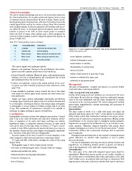
Figure 30.4: CT scan, saggital reconstruction. Slide shows retropulsed thoracic
B Sensory only Sensory sparing vertebra into spinal canal.
C Motor useless Motor sparing of no functional value
D Motor useful Motor sparing of functional value • acute kyphotic angulation;
E Recovery No functional deficit • widened interspinous space;
• axial rotation of vertebra;
The various spinal cord syndromes include: • discontinuity in contour lines;
• Anterior cord syndrome: Damage to the spinothalamic and cortico- • abnormal joints;
spinal tracts with resultant predominant motor weakness.
• atlanto-dental interval of more than 5 mm;
• Brown–Sequard’s syndrome: Hemicord injury with ipsilateral motor
weakness and loss of proprioception and contralateral loss of pain • narrow or widened disc space; and
and temperature below the level of injury.
• widening of apophyseal joints.
• Central cord syndrome: Injury to the central portions of the cervi- Management
cal spinal cord with resultant predominant motor affectation of the
upper limb. The goal of management of spinal cord injuries is to prevent further
injury and reduce neurological deficits.
• Conus medullaris syndrome: Injury towards the end of the spinal Initial management and evaluation
cord results in a mixed upper motor neurone and lower motor neu-
rone dysfunction. Ideally, initial management and evaluation are commenced at the scene
of the injury. In most African settings, however, prehospital manage-
Spinal cord injury without radiographic abnormality (SCIWORA) ment is not well established, and the initial management is usually
is a unique type of spinal cord injury common to children characterised commenced at the receiving hospital. The initial management includes
by posttraumatic neurological deficits with normal plain radiographs resuscitation, immobilisation, constant monitoring, and assessment of
or tomographs. It occurs mostly in children younger than 8–10 years the injured child. 24
of age. The mechanism of occurrence is thought to be vascular or Resuscitation
ischaemic in origin, resulting in spinal cord infarction.
The main causes of death of in a child with spinal cord injury are
Investigations aspiration and shock, and so the “ABC” of life support is commenced.
Radiographic evaluation is done after adequate resuscitation. A lateral Early airway control with endotracheal intubation and oxygen admin-
plain x-ray is the most informative and may show fractures, sublux- istration may be indicated in respiratory insufficiency. Manual in-line
ation, or angulation of the spine. Soft tissue swellings may indicate immobilisation of the cervical spine is mandatory during intubation.
ligamentous injury. In suspected odontoid fractures, an open mouth Hypotension accompanied by bradycardia may be present due to auto-
view can be done for the older child. In infants, a CT scan is recom- nomic paralysis. Therefore, adequate hydration with systolic blood
mended. At least 75% of patients with spinal cord injury have injury to pressure maintained at or above 90 mm Hg prevents shock. Volume
25
the vertebral column and thus some degree of radiographic abnormali- resuscitation suffices, but occasionally ionotropes such as ephedrine
ties. Therefore, initial plain films are indispensable. may be indicated.
Dynamic studies can be done to search for occult instability in the Nasogastric tube decompression of the stomach is instituted because
older cooperative child with neck pain but no neurologic deficit. gastric distention can interfere with respiration or lead to gastric
CT scans and MRI could further elucidate the extent of the injury mucosal ulceration.
(Figure 30.4). The loss of sympathetic tone may also lead to urinary retention and
Radiographic signs of cervical spine trauma include: hypothermia. An indwelling urethral catheter is passed, and attention
• soft tissue in retropharyngeal space >22 mm ( child not crying); paid to the temperature of the child with constant monitoring.
Immobilisation
• displaced prevertebral fat stripe;
The entire spine of the child with suspected spinal injury should be
• tracheal deviation and laryngeal dislocation; immobilised. Whole-body braces usually are not readily available, so
the cervical spine is immobilised with collars, particularly in the older
• vertebral malalignment;
child. Infants can be immobilised with sand bags or intravenous fluid
• loss of lordosis; bags secured at both sides of the head, with the head taped to the board.