
Page 2 - 63 craniocerebral-and-spinal-trauma30-35_opt
P. 2
Craniocerebral and Spinal Trauma 191
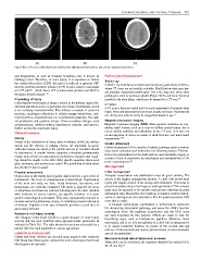
(A) (B) (C)
Figure 30.2: CT scans of (A) depressed skull fracture; (B) epidural haematoma; (C) chronic subdural haematoma.
and bradycardia, as well as irregular breathing—this is known as Radiological Assessment
Cushing’s triad. Therefore, in head injury, it is important to follow Skull x-ray
the cerebral blood flow (CBF). Because it is difficult to measure CBF
A skull x-ray is useful as an initial assessment tool, particularly in Africa,
directly, cerebral perfusion pressure (CPP) is used, which is calculated
where CT scans are not readily available. Skull fracture sites may her-
as: CPP = ICP – MAP, where ICP is intracranial pressure and MAP is
ald potential intracranial pathologies. The x-ray may also show other
the mean arterial pressure. 3,5
pathologies, such as pneumocephalus (Figure 30.3), and linear fractures
Physiology of injury parallel to the slice plane, which may be missed by a CT scan.
7,8
Following the initial injury at impact, known as the primary injury, bio- CT scan
chemical alterations occur, in particular, the release of glutamate, which A CT scan is the most useful tool for acute assessment of traumatic head
is an excitatory neurotransmitter. This initiates a cascade of cytotoxic injury. Bony and parenchymal lesions are usually well seen. Haematomas
reactions, resulting in alterations in cellular energy metabolism, cere- are clearly seen and can easily be categorised based on age. 9
bral blood flow, transmembrane ion concentration gradients, free radi-
cal production, and cytokine release. Gross secondary changes, such Magnetic resonance imaging
as haematomas, cerebral oedema, hypotension, seizures, and hypoxia, Magnetic resonance imaging (MRI) offers superior resolution in visu-
further worsen the neurologic injury. alising small lesions, such as is seen in diffuse axonal injury, but is
Clinical Features not as widely available and affordable as the CT scan. It is also not
an investigation of choice in terms of skull fractures and intracranial
History haematomas. 10,11
Details of the mechanism of injury, such as distance of fall, the surface Cranial ultrasound
struck, and the velocity of striking objects, are important. In motor
Cranial ultrasound (US) is usually a bedside technique used to monitor
vehicular trauma, the speed of the vehicle and use of restraints should
intracranial collections and ventricular size following trauma. This use-
be determined. A careful history regarding immediate posttraumatic
ful tool is underutilised for the child with an open fontanelle, largely as
events, such as loss of consciousness, its duration, seizures, and vomit-
a result of lack of experience by radiologists and unavailability of US
ing, should be sought. In the older child, specific questions about neck
to the neurosurgeons. 10
pain, numbness, and weakness are asked. The possibility of child abuse
should also be kept in mind. Management
Physical assessment Initial management
Observation of the mildly head injured child provides a great deal of Adequate resuscitation and stabilisation must be given priority. The
information. The level of consciousness is determined. Examination airway is the highest management priority. A child with severe head
of the head and scalp are done. Scalp abrasions, lacerations, and injury will require control of the airway with intubation. This helps to
12
haematomas are carefully examined. The skull is palpated for areas prevent secondary injury from hypoxia and hypercarbia. The cervical
of tenderness and fractures without inflicting pain. In older children spine must be assumed to be unstable until proven otherwise by plain
with moderate to severe injuries, age-specific behavior is a great radiographs later. Meanwhile, the breathing, circulation, and the stabili-
guide to neurological assessment. They may appropriately respond to sation of vital signs are then attended to. It is the postresuscitation GCS
noxious stimuli by grimacing, crying, or exhibiting a facial expression score that is useful.
of distress. Palpation of an open fontanelle provides a good idea of A focused neurological examination is performed to determine
intracranial pressure. life-threatening intracranial pathology and assess the child’s baseline
neurological level; the papillary examination and the GCS score
Assessment of injury severity
are most important for this purpose. Efforts are made to look for
The Glasgow Coma Scale (GCS) is a good measure of acute injury
lateralising signs, such as hemiparesis, pupillary dilatation, facial
severity and has been modified using age-appropriate parameters as
nerve palsy, and so on. The next priority in a child who is unresponsive
indicated in Table 30.1. The table shows the best score achievable by a
is to assess brainstem function by means of the corneal and gag
normal child for each parameter at various age groups.
reflexes. Corticosteroids and routine administration of anticonvulsants
Laboratory Assessment are not recommended.
Infants and small children can develop acute anaemia with relatively Measures to treat raised intracranial pressure
little blood loss. Haemogram and baseline serum electrolytes levels and Where ICP can be monitored, the treatment threshold for raised ICP is
blood gasses are assessed.
20–25 mm Hg.
13