
Page 16 - 58peadiatric-surgery-speciality1-7_opt
P. 16
18 Cardiovascular Physiology And Support
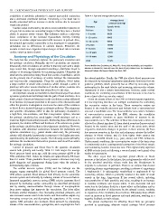
availability of metabolic substrates to support myocardial contraction Table 4.1: Age and average blood pressures.
and a decreased ventricular preload. Conversely, a low heart rate is Average blood
usually associated with an increase in stroke volume due to increased Age pressure (mm Hg)
ventricular preload.
Cardiac output is affected by the above-noted variables in patients of Premature Systolic 40–60
all ages, but neonates are somewhat unique in that they have a limited Full term 75/50
ability to increase stroke volume. The difference reflects a relatively 1–6 months 80/50
lower compliance of the neonatal myocardium, thereby limiting 6–12 months 90/65
increases in cardiac output associated with increases in preload. Also, 12–24 months 95/65
the neonatal myocardial contractility is less responsive to sympathetic
stimulation due to differences in calcium transits. Therefore, the 2–6 years 100/60
neonate is much more dependent upon changes in heart rate to increase 6–12 years 110/60
cardiac output in times of need. 12–16 years 110/65
Anatomy and Physiology of the Circulation 16–18 years 120/65
The body has two circulatory systems: the pulmonary circulation and Adult 125/75
the systemic circulation. Normally, the two circulations are separate.
Structurally, both circulations are similar in that they start with a single Source: Modified from Zuckerberg AL, Wetzel RC, Shock, fluid resuscitation, and coagulation
large vessel that, through sequential branchings, distributes blood to disorders. In Nichols DG, Yaster M, Lappe DG, Buck JR (eds). Golden Hour: The Handbook of
Advanced Pediatric Life Support. Mosby-Year Book, 1991.
arteries of decreasing diameter but increasing number. Ultimately the
small arteries (arterioles) empty blood into a series of capillaries, which
are the primary site of exchange of solutes between the intravascular the adrenal medullae. Finally, the CNS also affects blood pressure over
and extravascular compartments. From the capillaries, blood enters the long term by releasing vasopression (antidiuretic hormone) from the
a large number of small veins (venules), which, through a series of posterior pituitary, which primarily has an effect by increasing water
junctions with other venous structures of similar calibre, coalesce into reabsorption by the renal tubules and increasing intravascular volume.
several large venous structures that return blood to the atria. Vasopressin is also a potent vasoconstrictor; however, under normal
An important concept to remember is that blood always flows down conditions, the circulating concentration of this hormone is too low to
a pressure gradient and will always take the path of least resistance. As a have a direct effect on vascular control.
result, points of increased resistance in the circulatory system will result Given the potent effects of the CNS on blood pressure regulation,
in an increase in pressure proximal to the point of the obstruction until it is not surprising that there are multiple mechanisms for controlling
either the pressure is adequate to overcome the cause of the resistance the vasomotor centres in the brain. These vasomotor centres are
or blood flow is diverted through an alternate pathway. The pulmonary located in the medulla and pons and include both a vasoconstrictor
and systemic circulations differ in that the pulmonary circulation is area and a vasodilator area. The vasoconstrictor area causes excitation
a high-flow, low-resistance, and thus low-pressure system, whereas of vasoconstrictor neurons in the SNS, whereas the vasodilator
the systemic circulation has much higher overall resistance and as a centre primarily functions to cause inhibition of neurons in the
result has higher intraluminal pressures. Reflecting these differences in vasoconstrictor area. The activities of these two vasomotor centres are
pressures, the relative stiffness and thickness of the arteries are greater affected by afferent impulses (1) from stretch receptors (baroreceptors)
in the systemic circulation than in the pulmonary circulation. However, located in the carotid sinus and the wall of the aortic arch, which
in patients with abnormal connections between the pulmonary and respond to short-term changes in pressure in these arteries; (2) from
systemic circulations (e.g., patent ductus arteriosus), the pulmonary low pressure receptors in the atria and pulmonary arteries that reflect
arteries will ultimately hypertrophy in response to the higher pressures changes in blood volume; and (3) from higher brain centres that
experienced by the pulmonary vessels. The following section primarily respond to stressful stimuli (e.g., pain, alarm) and CNS ischaemia.
addresses the mechanisms that control pressure and blood flow within Under normal conditions, the vasoconstrictor centre of the brain stem
the systemic circulation. is continuously active, causing partial contraction of the blood vessels
Control of pressure and blood flow in the systemic circulation and maintaining baseline vasomotor tone. This explains why rapid loss
occurs both globally and locally. Table 4.1 lists ranges for average of SNS activity (such as following a cervical spinal cord injury) often
blood pressure based upon age, with the mean pressure +20% at the results in hypotension.
95% confidence limit. Values for females are approximately 5% lower The other organ that has important global effects on blood
than for males. These paediatric blood pressure references may help pressure is the kidney. In the kidney, the juxtaglomerular cells located
guide diagnosis and management during times when the patient is in the proximal arterioles release renin into the bloodstream in
demonstrating signs of shock. response to a decrease in perfusion. Renin is an enzyme that cleaves
The central nervous system (CNS) and the kidneys are the circulating plasma angiotensinogen, resulting in release of angiotensin
primary organs responsible for global blood pressure control. The I. Angiotensin I is subsequently metabolised to angiotensin II by
CNS affects systemic blood pressure both directly via the autonomic converting enzyme, which is primarily located in the walls of small
nervous system and indirectly by inducing release of humoral factors. vessels in the lung. Angiotensin II has several effects, including
Increased sympathetic nervous system (SNS) activity increases blood vasoconstriction of both arterioles and veins, resulting in an increase
pressure by increasing cardiac contractility (as described previously) in vascular resistance and venous return. It also decreases salt and
and by causing vasoconstriction through release of norepinephrine water loss by the kidney (both by a direct effect on the kidney and by
from nerve endings that innervate the vasculature. The latter effect stimulating secretion of aldosterone by the adrenal cortex), resulting
increases arterial resistance, thus increasing proximal pressures, and in expansion of the circulating blood volume. Ultimately, these effects
also increases venous return to the heart—and thus cardiac output—by cause an increase in blood pressure and renal perfusion, resulting in a
increasing venous tone and decreasing the capacitance of the venous negative feedback on renin release.
system. SNS activation also increases blood pressure by stimulating The global mechanisms for affecting blood flow are primarily
release of the vasoconstrictors, epinephrine, and norepinephrine from involved in maintaining adequate central systemic blood pressure