
Page 102 - 85 cell signalling pathways
P. 102
Cell Signalling Biology Michael J. Berridge Module 2 Cell Signalling Pathways 2 102
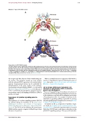
Module 2: Figure STAT1/DNA complex
Organization of a STAT1/DNA complex.
The ribbon diagram shown in (A) illustrates how the two DNA-binding domains of the two subunits attach the dimer to the DNA helix. The linker domain
(orange) attaches to the SH2 (Src homology 2) domains (light blue) that hold the molecule together through the intermolecular SH2--phosphotyrosine
interaction. The molecular surface representation shown in (B) has the same orientation as in (A). The local electrostatic potential over the cell surface
is represented by the colouring, with blue representing positive and red negative potentials. (Reproduced from Cell, Vol. 93, Chen, X., Vinkemeier,
U., Zhao, Y., Jeruzalmi, D., Darnell, Jr, J.E. and Kuriyan, J., Crystal structure of a tyrosine phosphorylated STAT-1 dimer bound to DNA, pp. 827--839.
Copyright (1998), with permission from Elsevier; see Chen et al. 1998.
the receptor and then dimerize before translocating into There is a marked increase in expression of the SOCS-3
the nucleus. This activation cascade can occur quickly, isoform in hypothalamic neurons following the action of
with the activated STATs appearing in the nucleus within leptin during the control of food intake and body weight
minutes. Transcriptional activity can be modulated by (Module 7: control of food intake).
phosphorylation of the serine residue in the transcrip-
tional activation domain (TAD) of STAT 1, 3, 4, 5A and 5B Janus kinase (JAK)/signal transducer and
(Module 2: Figure JAK and STAT structure). In the case of activator of transcription (STAT) function in
growth and development
STAT 1 and STAT4, phosphorylation enhances transcrip-
The Janus kinase (JAK)/signal transducer and activator of
tional activity, whereas the binding of STAT5a to DNA is
transcription (STAT) signalling pathway has a primary role
greatly prolonged.
in the regulation of growth and development, particularly
Suppressor of cytokine signalling proteins of haematopoietic cells. For example, it functions in the
(SOCS) interleukin-2 signalling pathway responsible for driving
The suppressor of cytokine signalling proteins (SOCSs) the final signalling steps in lymphocyte activation (Module
are induced during the activation of the Janus kinase 9: Figure T cell signalling map).
(JAK)/signal transducer and activator of transcription Amutationinthe γ c component of cytokine recept-
(STAT) signalling pathway (Module 2: Figure JAK/STAT ors results in X-linked severe combined immune defi-
function). The SOCS act by inhibiting the Janus kinases ciency (X-SCID). Since JAK3 associates with γ c , it seemed
(JAKs) and thus operate a negative-feedback loop to limit likely that X-SCID may result from an alteration in the
the action of cytokines. JAK/STAT signalling pathway. This was confirmed when
C 2012 Portland Press Limited www.cellsignallingbiology.org