
Page 127 - LECTURE NOTES
P. 127
Note: If too intense a light source is used the contrast will not be adequate
and the unstained fungi will not be seen.
Dermatophytes in skin scales: look for branching septate hyphae with angular or
spherical arthrospores, usually in chains. All species of ringworm fungi have a similar
appearance
Fungi need to be distinguished from epidermal cell outlines, elastic fibers, and artifacts
such as intracellular cholesterol (mosaic fungus) and strands of cotton or vegetable
fibers. Ringworm fungal hyphae can be differentiated from these structures by their
branching, uniform width, and cross- walls (septa), which can be seen when using 40-x
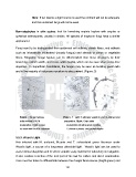
objective. In Superficial Candidiasis, the fungus may be seen as budding yeast cells
and in the majority of instances mycelium is also present. (Figure: 3)
Figure; 3 fungal hyphae Figure: 4 Left; C.albicans yeasts in wet un stained and
arthroconida in KOH preparation. Right: Gram stain
preparation of skin scales preparation showing gram positive
as seen with the 40x objective C albicans yeasts and psuedohyphe
5.5.3. Wood’s Light
Hair infected with M. audounii, M.canis and T. schoenleinii green fluoresce under
Wood’s light, a source of a long-wave ultraviolet light. Wood’s light can be used to
assist clinical diagnosis and to select suitable scalp material for laboratory investigation.
It also enables selection of the best part of the hair for culture and direct examination.
Care must be taken to differentiate between true fungal fluorescence (bright green) and
121