
Page 5 - PII: S0044-8486(99)00371-3
P. 5
)
D. Lemos et al.rAquaculture 186 (2000 89–105 93
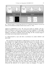
Fig. 1. Schematic sequence of substrate-SDS-PAGE protocols for determining Ž. a proteinase composition and
activity, and Ž. b proteinaceous proteinase inhibitors Garcia-Carreno et al., 1993 .
˜
.
Ž
separated by SDS-PAGE as above. The twin gel is soaked for 30 min at 58Cina
solution containing the target enzyme, a commercial enzyme preparation or a hepatopan-
creas extract. Then, it is washed with distilled water and soaked in a solution of the
substrate as described for proteinase activity assay ŽFig. 1b . The molecular weight of
.
active bands can be determined as described above.
2.3. Characterization of type and class of proteinases by specific inhibitors and
SDS-PAGE
Most proteinases from the digestive system belong to the serine class ŽEC 3.4.21.x ,
.
the exception is the stomach proteinase pepsin that belongs to the aspartic class. To
determine the class and specificity of the enzyme, the proteinase under study is
incubated with specific inhibitors ŽTable 1 for serine class, trypsin or chymotrypsin
.
ŽGarcia-Carreno and Haard, 1993 . Solutions of 20 mM tosyl-lysine chloromethyl
˜
.
ketone ŽTLCK in 1 mM HCl, 200 mM phenylmethylsulphonyl fluoride PMSF in
.
Ž
.
2-propanol, or 0.5 mM carbobenzoxy-phenylalanine chloromethyl ketone ŽZPCK in 50
.
mM phosphate buffer, pH 7.8, are individually mixed with the enzyme extracts in a ratio
of 1:10 Žinhibitorrextract and incubated for 1 h at 258C. Distilled water is used instead
.
of inhibitors in control. Then the inhibitor–enzyme mixture is diluted with the sample
buffer and loaded onto the gel as described above. After electrophoresis, lanes are cut
apart for molecular weight standard and total protein lanes, and activity and inhibition
lanes. The former lane is immediately stained. Activity and inhibition lanes are
immersed in 3% casein following the described procedure for activity. Bands with
proteinase inhibitors are compared with control proteinase lanes Žno inhibition to
.
identify the inhibitory effect on active bands. Since TLCK, ZPCK, and PMSF are
specific trypsin, chymotrypsin and serine proteinase inhibitors ŽTable 1 , those bands
.
affected by the inhibitors will be smaller or may even disappear. The degree of