
Page 89 - 84 human physiolofy part-1
P. 89
Senses
Retina
The third or the innermost layer of the eye is call the retina. In adult humans the entire retina is
72% of a sphere about 22 mm in diameter. The retina lays over the back two thirds of the choroid
coat, which is located in the posterior compartment. The compartment is filled with vitreous
humor which is a clear, gelatinous material. Within the retina there are cells called rod cells and
cone cells also known as photoreceptors. The rod cells are very sensitive to light and do not see
color, that is why when we are in a darkened room we see only shades of gray. The cone cells are
sensitive to different wavelengths of light, and that is how we are able to tell different colors. It is
a lack of cones sensitive to red, blue, or green light that causes individuals to have deficiencies in
color vision or various kinds of color blindness. At the center of the retina is the optic disc,
sometimes known as "the blind spot" because it lacks photoreceptors. It is where the optic nerve
leaves the eye and takes the nerve impulses to the brain. The cornea and the lens of the eye
focuses the light onto a small area of the retina called the fovea centralis where the cone cells are
densely packed. The fovea is a pit that has the highest visual acuity and is responsible for our
sharp central vision - there are no rods in the fovea.
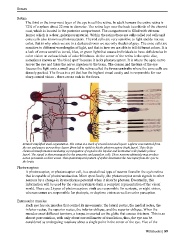
Retina's simplified axial organization. The retina is a stack of several neuronal layers. Light is concentrated from
the eye and passes across these layers (from left to right) to hit the photoreceptors (right layer). This elicits
chemical transformation mediating a propagation of signal to the bipolar and horizontal cells (middle yellow
layer). The signal is then propagated to the amacrine and ganglion cells. These neurons ultimately may produce
action potentials on their axons. This spatiotemporal pattern of spikes determines the raw input from the eyes to
the brain.
Photoreceptors
A photoreceptor, or photoreceptor cell, is a specialized type of neuron found in the eye's retina
that is capable of phototransduction. More specifically, the photoreceptor sends signals to other
neurons by a change in its membrane potential when it absorbs photons. Eventually, this
information will be used by the visual system to form a complete representation of the visual
world. There are 2 types of photoreceptors: rods are responsible for scotopic, or night vision,
whereas cones are responsible for photopic, or daytime vision as well as color perception.
Extraocular muscles
Each eye has six muscles that control its movements: the lateral rectus, the medial rectus, the
inferior rectus, the superior rectus, the inferior oblique, and the superior oblique. When the
muscles exert different tensions, a torque is exerted on the globe that causes it to turn. This is an
almost pure rotation, with only about one millimeter of translation, thus, the eye can be
considered as undergoing rotations about a single point in the center of the eye. Five of the
Wikibooks | 89