
Page 22 - 68 stomac-duodenum-&-small-intestine59-65_opt
P. 22
CHAPTER 64
Vitelline Duct Anomalies
Bankole S. Rouma
Kokila Lakhoo
Introduction Demographics
Vitelline duct or omphalomesenteric duct anomalies are secondary The most frequent malformation is Meckel’s diverticulum, with an
to the persistence of the embryonic vitelline duct, which normally incidence of 2–3% of the population, but it is one of the most unlikely
obliterates by weeks 5–9 of intrauterine life. These anomalies occur in to cause symptoms. About 4% of children with a Meckel’s diverticulum
approximately 2% of the population and may remain silent throughout develop symptoms, and more than 60% of those who develop symp-
2–5
life, or may present incidentally sometimes with an intraabdominal com- toms are younger than 2 years of age. The male-to-female complica-
plication. Although Meckel’s diverticulum is the most common vitelline tion rate ratio is about 3:1. 3
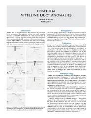
duct anomaly (Figure 64.1(G)), a patent vitelline duct (Figure 64.1(A)) Embryology
is the most common symptomatic presentation in developing countries. 1
During week 3 of gestation, the midgut is open into the yolk sac, which
does not grow as rapidly as the rest of the embryo. Subsequently, by
week 5, the connection with the yolk sac becomes narrowed and is
then termed a yolk stalk, vitelline duct, or omphalomesenteric duct.
Normally, the vitelline duct disappears by gestational week 9, just
before the midgut returns to the abdomen. Persistence of some portion
of the vitelline duct results in a number of congenital anomalies, of
which Meckel’s diverticulum is the most common. This anomaly is
variable in length and location, but most often it is observed as a 1–5
cm intestinal diverticulum projecting from the antimesenteric wall of
the ileum within 100 cm of the caecum. It possesses all three layers
of the intestinal wall and has its own blood supply. The connection in
a patent vitelline duct is usually to the ileum, but less commonly may
be to the appendix or colon. In other cases, part of the vitelline duct
1
within the abdominal wall persists, forming an open omphalomes-
enteric fistula, an enterocyst, or a fibrous band connecting the small
bowel to the umbilicus. 2–7
Pathophysiology
Vitelline duct malformations comprise a wide spectrum of anatomic
structures, depending on the degree of involution of the vitelline duct.
The most common anomaly is Meckel’s diverticulum, described as
being 60 cm from the ileocaecal valve, 2 cm in diameter, 3 cm in length,
and not attached to the abdominal wall. Most complications of these
abnormalities are related to ectopic tissue (gastric, pancreatic, colonic,
endometriosis, or hepatobiliary). 7
Ectopic gastric tissue usually causes bleeding from ulceration of
the adjacent ileal mucosa. The ileal mucosa is not equipped to buffer
the acid produced by the ectopic gastric mucosa and thus is prone to
ulceration. The site of the ulceration is most often at the junction of the
normal ileal mucosa and the ectopic gastric mucosa. Some studies have
shown a very low colonisation rate with Helicobacter pylori in children
with ulcerative bleeding of Meckel’s diverticulum. 3
Intestinal obstruction may be caused by a Meckel’s diverticulum
attached to the umbilicus by a fibrous cord or by a fibrous cord between
the ileum and the umbilicus. This may lead to a volvulus around
the fibrous cord. A persistent vitelline artery, an end artery from the
superior mesenteric artery, may cause obstruction and volvulus. Bowel
Figure 64.1: Remnants of the omphalomesenteric duct: (A) patent vitelline duct; (B) obstruction can also occur by intussusception with the diverticulum
patent vitelline duct covered by skin; (C) Meckel’s diverticulum with fibrous cord; as a lead point or by herniation or prolapse of the bowel through a
(D) cyst with fibrous cord; (E) cyst; (F) fibrous cord; (G) Meckel’s diverticulum. patent omphalomesenteric fistula (with a characteristic “ram’s horn”
appearance). Obstruction may be caused by phytobezoar. 6,7
5