
Page 25 - 62 paediatric-trama25-29_opt
P. 25
Thoracic Trauma 181
Management is symptomatic, but intensive care is often required
in the initial phase, where there is danger of respiratory collapse and
ventilation might be indicated for adequate oxygenation. The prognosis
is good if infection does not occur; healing can be expected within 1–2
weeks. Unfortunately, in two-thirds of these cases, infection occurs
due to the extravasation of fluid and blood in interstitium and alveoli,
which creates an excellent microbial culture medium. Ventilation
efforts are often poor due to pain, and without active and passive chest
physiotherapy, the prognosis is poor.
Pulmonary haematoma is rare. It is usually caused by an injury to a
major blood vessel within the lung, creating a so-called coin-lesion in
the lung tissue. Management is nonoperative, except in massive bleeds.
Simple Pneumothorax
Pneumothorax is a common occurrence in childhood chest injury.
Collapse of the lung might be caused by a penetrating injury, a rupture
of lung parenchyma, or a tear in the oesophagus or tracheobronchial tree.
Physical signs are diminished breath sounds, poor motion of the
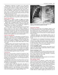
hemithorax, hyperresonance to percussion, subcutaneous emphysema, Figure 28.1: Chest radiograph of a ruptured left hemidiaphragm, with
and deviation of the trachea to the ipsilateral site. Diagnosis is displacement of the heart and mediastinum to the right.
confirmed with an erect expiratory chest radiograph.
Treatment consists of a tube thoracostomy in the 4th intercostal
space, in the anterior axillary line, under adequate analgesia. Care Oesophageal Injuries
should be taken not to cause injury to the lung parenchyma or Fortunately, due to the location of the oesophagus, injuries to it are
diaphragm during the insertion of the tube. An underwater seal should rare. Transmitted pressure from the stomach may cause either Mallory-
immediately be connected to the bottle. If the child is asymptomatic and Weiss bleeding (if the lower oesophageal sphincter is closed) or the
can be closely monitored, aspiration or even observation of a simple more sinister Boerhaave syndrome, characterised by perforation of the
pneumothorax may be appropriate, but the resources to rapidly insert a lower oesophagus into the left chest cavity (if the upper oesophageal
chest tube must be available in the event of any deterioration. 5 sphincter is closed).
Penetrating injuries may cause oesophageal injuries if they are
Tension Pneumothorax
transthoracic. Radiographic contrast studies and/or endoscopies are
Progressive accumulation of air under pressure in the pleural space is
strongly advocated in these cases. A nonionic contrast material should
usually due to a valve-effect tear in the lung parenchyma. It may lead
be used.
to ipsilateral collapse of the lung and mediastinal shift, thereby com-
The management of the oesophageal injuries depends on the nature
pressing the (only properly ventilating) contralateral lung. This might
of the injury, the timing of presentation, and the location. With the
result in severe impairment of ventilation as well as compromise the
exception of major (high-velocity) gunshot injuries, the majority can
venous return to the heart, and is often a lethal condition if not acted
be repaired primarily within 24 hours of the injury. Beyond the first
upon rapidly.
24 hours, the operative strategy may include oesophageal diversion,
Diagnosis should be made clinically. Decreased breathing sounds,
exclusion, T-tube drainage, or even total oesophagectomy.
a hyperinflated ipsilateral hemithorax, trachea deviation to the
contralateral side, and a severely distressed patient all indicate that Cervical oesophageal injuries
a fast needle-puncture of the anterior chest (2nd intercostal space, Cervical oesophageal injuries rarely represent a large problem because
midclavicular line) will be life saving. The needle has to be replaced leakage from a repair produces localised tissue infection or abscess,
by a proper tube thoracostomy as soon as possible because blockage which can be drained externally.
occurs frequently, and the excursions of an inflated lung will damage its Thoracic oesophageal injuries
visceral pleural surface against the sharp tip of the needle. Thoracic oesophageal injuries are notorious for the fast spread of sali-
Haemothorax va, food, and acid from the stomach through the injury into the chest,
Haemothorax is the accumulation of blood in the pleural space. Up to able to cause a rampant and usual lethal mediastinitis. Oesophageal
40% of the blood volume can easily be lost in one pleural cavity. The diversion might be indicated in these cases.
blood loss usually arises from injury to a major artery, either from the Abdominal oesophageal injuries
chest wall or the lung, although this is not always the case. Persistent Abdominal oesophageal injuries will usual present as an acute abdomen
bleeding from an intercostal artery or a tear in the lung parenchyma can and will require a laparotomy for repair.
also produce major blood loss. Diaphragmatic Injuries
The diagnosis is made clinically and confirmed with an erect chest
radiograph. Blood in the lower part of the pleural cavity often causes Traumatic disruption of the diaphragm is usually caused by blunt
referred pain in the upper abdomen. Once the haemothorax is drained, trauma. It involves the left side in the majority of cases. The injury is
the abdominal symptoms disappear. high velocity in nature, such as from motor vehicle collisions and falls
Treatment consists of chest tube thoracostomy; only rarely is a from a height. Because the force required to damage the diaphragm is
thoracotomy indicated. The main indications for thoracotomy are considerable, associated injuries are common (about 80%) and include
ongoing active bleed while an intercostal drain is in place, or an infected intrathoracic and intraabdominal as well as extratruncal injuries.
haemothorax (usually 5–7 days after injury). On rare occasions, a The clinical presentation varies according to the associated injuries;
massive haemothorax may lead to a tension haemothorax with deviation an isolated diaphragmatic rupture can easily be misdiagnosed. In
of the heart and mediastinum to the opposite side (Figure 28.1). children, the mechanism of injury might be slightly different from that
in adults. Whereas in adults the typical injury involves the dome of