
Page 2 - 61 surgical-infection&infestations20-24_opt
P. 2
Omphalitis 125
Clinical Presentation Medical Treatment
Local signs of omphalitis include purulent or foul-smelling discharge Treatment of uncomplicated cases requires prompt antibiotic therapy.
from the umbilicus/umbilical stump, periumbilical erythema, oedaema, Antibiotics are the mainstay of medical treatment of omphalitis. Antibiotics
and tenderness. Systemic signs include fever (temperature >38°C) specifically active against Staphylococcus aureus and an aminoglycoside
or hypothermia (temperature <36°C), unstable temperature, or jaun- to cover for both gram-positive and gram-negative organisms are used.
dice. Other systemic manifestations may include tachycardia (heart The local antibiotic susceptibility patterns need to be considered in the
rate >180/min), hypotension and delayed capillary refill, tachypnoea initial therapy. Examples include ampiclox, cloxacillin, flucloxacillin, and
(respiratory rate >60/min), signs of respiratory distress or apnoea, or methicillin in combination with gentamycin. Metronidazole may be added
abdominal distention with absent bowel sounds. Central nervous sys- when anaerobes are suspected. Duration of treatment is typically for 10–14
tem involvement may manifest as irritability, lethargy, poor suckling, days with initial parenteral therapy for complicated cases. A short antibiotic
hypotonia, or hypertonia. A history of delayed cord separation may be therapy of 7 days is adequate for simple uncomplicated omphalitis.
present in LAD syndrome. Complications such as respiratory failure, hypotension, and
In advanced cases, the infant may present with septic shock or disseminated intravascular coagulation (DIC) arising from infection may
necrotising fasciitis (NF). NF is a severe complication of omphalitis require supportive care in the form of intravenous fluids, fresh whole
that should be considered if the local signs have progressed to include blood, fresh frozen plasma, platelets, or cryoprecipitate.
a peau d’orange appearance, discolouration or bruising of the skin, skin Treatment of Surgical Complications
necrosis, and crepitation.
The surgical complications of omphalitis could be acute/early or long term/
Differential Diagnoses late and tend to be associated with significant morbidity and mortality. In
The differential diagnoses of omphalitis (and specific features of each) addition to medical treatment for ongoing/active omphalitis, the surgical
include: treatment is handled according to the surgical complication.
Necrotising Fasciitis
• umbilical granuloma (visible granuloma at the umbilicus);
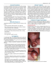
Necrotising fasciitis is one of the most commonly reported serious com-
• patent vitello-intestinal duct remnants (cystic swelling or fistulous plications of omphalitis, 1,8–12 occurring in 26% of patients with major
opening with feculent matter discharging); complications, according to one report. It has been noted to occur in 13.5%
6
8
• patent urachus (fistulous opening with urine discharging) or urachal cyst; of neonates with omphalitis. The condition starts initially as periumbilical
cellulitis, which, without treatment, progresses rapidly to necrosis of the
• necrotising enterocolitis (abdominal distention, bilious vomiting, skin and subcutaneous tissue (Figures 20.2 and 20.3), and in some instances,
bloody stools);
• general sepsis; and
• rarely, appendiculo-omphalic anomalies.
Investigations
A microbiological swab of the umbilicus should be sent for aerobic and
anaerobic cultures. A blood culture should be included when appropriate. A
blood count with differential for white cell counts may show a neutrohilia
(or occasionally a neutropaenia).
Other investigations are necessary either to rule out other differential
diagnoses or to diagnose complications. Diagnostics may include the
following:
• A plain abdominal radiograph is useful if necrotising enterocolitis is
suspected. In addition, it may reveal intraperitoneal gas in those with
peritonitis (caused by gas-producing bacteria). Multiple fluid levels
may suggest adhesion obstruction but may also be present in simple
ileus. Gas may be present within the subcutaneous tissue of the abdom-
inal wall when clostridial infection is involved.
• Abdominal ultrasonography is useful in imaging the abdominal wall if
a cyst is suspected It is helpful in the diagnosis of intraperitoneal, retro-
peritoneal, and hepatic abscesses.
• Dopplar ultrasonography is helpful if portal vein thrombosis is
suspected.
• A fistulogram is indicated if a fistulous connection to the umbilicus is
discovered. This will help define the anatomy of a vitello-intestinal or
urachal remnant.
• Rarely, magnetic resonance imaging (MRI) or a computed tomog-
raphy (CT) scan may be useful in assessing or ruling out congenital
tracts or fistulas. Also rarely, a CT scan may be necessary to adequately Figure 20.2: Periumbilical cellulitis with early necrosis of scrotal skin.
localise intraabdominal abscesses in difficult diagnostic cases.