
Page 2 - 59peadiatric-surgery-speciality8-14_opt
P. 2
Wound Healing 41
vasoconstriction at the site of injury is followed by vasodilatation,
which increases local blood flow to the area. Vascular permeability is
increased through activation of the complement pathways and coagula-
tion cascade. There is an influx of cells and substrates necessary for
healing, including early neutrophil scavengers, plasma proteins, and
activated complement fragments.
A predominance of neutrophils within the first 24 hours act to sterilise
the wound (Figure 8.3). After 2–3 days, the cell population shifts to a
predominance of macrophages derived from resident macrophages and
monocytes that are attracted to and infiltrate the wound. Macrophages
continue phagocytosis and secrete GFs and cytokines, which induce
fibroblast proliferation, angiogenesis, and production of extracellular
matrix. Lymphocytes begin to appear in small numbers, but little is
understood about their role in the wound-healing process.
Proliferation
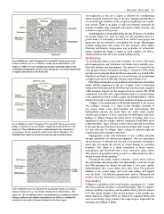
Source: Modified from Cohen IK, Diegelmann RF, Crossland MC. Wound care and wound The proliferative phase begins with formation of a fibrin, fibronectin
healing. In: Schwartz SI, et al., eds. Principles of Surgery, 6th ed. McGraw-Hill Inc., 1994.
glycosaminoglycan, and hyaluronic acid matrix that is initially popu-
Figure 8.3: Within 24 hours following tissue injury, neutrophils attach to the lated with platelets and macrophages. The various GFs secreted by the
endothelium (margination) and then move through the vessel walls (diapedesis) macrophages enhance fibroplasia, and there is migration of fibroblasts
to migrate (chemotaxis) to the wound site.
into the wound using the fibrin and fibronectin matrix as a scaffold. The
fibroblasts proliferate in response to GFs and become the predominant
cell type by the third to fifth day following injury (Figure 8.4).
Fibroblasts entering the wound proliferate and synthesize extracellular
matrix (ECM) components at the site of injury. There is interaction
between the fibroblasts and the ECM through transmembrane receptors
called integrins. Ligands for the integrin receptors include GFs, ECM
components, and other cells. Ligand binding leads to structural change
in the cytoplasmic domain of the receptor and phosphorylation. Signal
transduction leads to transcription factor synthesis and gene expression.
Collagen is the predominant ECM protein deposited at the wound.
The collagen molecule is a triple helical structure abundant in
two unique amino acids, hydroxyproline and hydroxylysine. The
hydroxylation process that forms these two amino acids requires
ascorbic acid (vitamin C) and is necessary for stabilisation and cross-
linking of collagen. During the initial phases of healing, there is an
8
abundance of type III collagen, which is composed of thin fibrils and is
Source: Modified from Cohen IK, Diegelmann RF, Crossland MC. Wound care and wound relatively pliable. Type I collagen is also formed, and with remodelling
healing. In: Schwartz SI, et al., eds. Principles of Surgery, 6th ed. McGraw-Hill Inc., 1994. it becomes the most abundant form found in normal adult wounds at a
Figure 8.4: The proliferation phase is characterised by the movement of 4:1 ratio with type III collagen. Type I collagen is relatively rigid and
macrophages into the wound site, which in turn attracts fibroblasts. The imparts high tensile strength to the tissue. 5
fibroblasts then repair the wound by producing new connective tissue.
Angiogenesis occurs with formation of new capillary networks
through endothelial cell division and migration. This new vasculature
allows delivery of nutrients and removal of by-products. Granulation
tissue may accompany the process in wound healing by secondary
intention. This tissue is a dense population of blood vessels,
macrophages, and fibroblasts with a loose connective tissue matrix.
The presence of granulation tissue is used as a clinical indicator that a
wound is ready for skin grafting. 9
Throughout this phase, wound contracture occurs, which leads to
the surrounding skin being pulled circumferentially toward the wound
bed. This decreases the wound size and helps it close more rapidly.
Epithelialisation also occurs within hours after injury. The epidermis
thickens at the wound edges, and basal cells enlarge and migrate
over the defect. Cell adhesion glycoproteins, such as fibronectin and
tenascin, form the framework to facilitate the epithelial cell migration.
Remodelling
Collagen accumulation in the wound reaches a maximum at 2–3 weeks
after injury, and the transition to remodelling begins. There is a balance
Source: Modified from Cohen IK, Diegelmann RF, Crossland MC. Wound care and wound between synthesis, deposition, and degradation during this time (Figure
healing. In: Schwartz SI, et al., eds. Principles of Surgery, 6th ed. McGraw-Hill Inc., 1994.
8.5). The tensile strength of the wound increases as the initially ran-
Figure 8.5: The remodelling phase is characterised by an equilibrium between domly deposited collagen fibrils are replaced by organised fibrils with
collagen synthesis and collagen degradation in an effort to re-establish the
connective tissue matrix that was destroyed by the tissue injury. more cross-linking. Lysyl oxidase is the major enzyme responsible for
ensuring cross-linking of fibrils.