
Page 34 - 58peadiatric-surgery-speciality1-7_opt
P. 34
36 Haemoglobinopathies
tration in the splenic remnant still exists, so this approach needs further Preliminary laboratory investigations are often unhelpful in
evaluation. In the meantime, splenectomy should generally be reserved distinguishing infection from infarction because both conditions
for selected patients with recurrent splenic sequestration crises or those can cause a leucocytosis with raised inflammatory markers. Blood
who develop red cell alloantibodies following transfusion therapy. cultures taken before the commencement of antibiotics can be
Gallstone Disease invaluable, as can culture of a bone or joint aspirate if there is
Pigment gallstones are a frequent complication of sickle cell disease evidence of fluid accumulation.
because continuous haemolysis leads to increased bilirubin excretion Imaging investigations are also confounded by the similarity
and subsequent stone formation. Although many children are asymp- between the radiographic appearances of bony infarction and infection.
tomatic, they can experience the full range of gallstone disease from Plain radiographs can be normal in the early stages of both conditions,
biliary colic to cholangitis. and the periositis and osteopaenia seen in acute osteomyelitis can also
Management of the acute complications of gallstones is the occur in vaso-occlusion. The imaging modality of choice for suspected
same as in the general population, and elective cholecystectomy osteomyelitis is magnetic resonance imaging (MRI), where it is
is recommended in patients with symptomatic cholelithiasis. The available, but even this is not 100% specific for differentiating infection
management of asymptomatic gallstones is less clear, but many from infarction. Ultrasonography, which is showing promise in the
would advocate cholecystectomy to avoid subsequent difficulty in diagnosis of osteomyelitis in children in particular, should be used to
distinguishing acute cholecystitis from vaso-occlusive painful episodes. guide any aspiration procedures.
The management of vaso-occlusive crises is largely supportive,
Orthopaedic Manifestations
focusing predominantly on pain management. By contrast, the first line
Bone-related symptoms are the most common reason for children with
management of osteomyelitis requires urgent parenteral antibiotics,
sickle cell disease to present to hospital. The osteoarticular manifesta-
ideally directed at whatever organism has been isolated. When
tions of sickle cell disease can be classified as acute or chronic, as
antibiotics are being started empirically, it is important to bear in
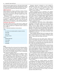
shown in Table 7.3.
mind that patients with SCD are more predisposed than the general
Table 7.3: Osteoarticular manifestations of sickle cell disease. population to contracting Salmonella osteomyelitis. Other organisms
that cause bone infection in this population include Staphylococcus
Acute
aureus, Haemophilus influenzae, and Escherichia coli. Third-generation
Vaso-occlusive crises (including dactylitis and diaphyseal infarction) cephalosporins are often used in this setting, and treatment should
continue for at least 6 weeks.
Osteomyelitis
Surgical drainage is generally believed to be required only in those
Septic arthritis cases of osteomyelitis that are not responding to antibiotics or where
there is evidence of abscess formation. However, the exact timing and
Pathological fractures
method of surgical intervention remains controversial.
Chronic
Chronic
Avascular osteonecrosis Avascular osteonecrosis is the most common chronic complication of
Chronic arthritis sickle cell bone disease and is believed to affect up to 41% of these
patients. It occurs when repeated bone infarction leads to destruction
Osteoporosis and breakdown of an area of bone, and it most often occurs at the
Osteomyelitis femoral head. Other areas affected include the head of the humerus, the
knee, and the small joints of the hands and feet.
Sufferers describe pain and limited movement at the affected joint;
Acute examination may reveal localised tenderness, with restriction of both
In a child presenting with acute bone pain, the most important distinc- active and passive joint movements. Initial investigations should
tion to make is between bone infarction and bone infection. Although include a plain radiograph, which may be diagnostic in more advanced
the vaso-occlusive crises that lead to bone infarction are up to 50 times cases, showing flattening or collapse of the articular surfaces and
more common than osteomyelitis, there is potential for extensive dam- subchondral radiolucency. Less advanced cases may show evidence of
age to the bone and surrounding structures as well as overwhelming sclerosis. MRI is the second-line investigation of choice.
sepsis if an infection remains untreated. When considering treatments for avascular necrosis in patients with
It is difficult to distinguish between the two conditions on clinical SCD, it is important to note the differences between this population
criteria alone because the archetypal features of osteomyelitis—namely, and nonsickle patients with the same condition. Not only is the
pain, swelling, and fever—are also common in vaso-occlusive crises. A pathophysiology of osteonecrosis in SCD thought to differ from that
history of a painful episode that has lasted longer than 1 to 2 weeks or of osteonecrosis from other causes, but the quality of the surrounding
pain in a distribution that does not conform to previous painful crises bone is often much poorer in this group of patients. Combined with
should raise suspicions of an alternative underlying cause. Infection their increased anaesthetic risk, this makes SCD patients less attractive
is not the only differential; stress fractures should also be considered. surgical candidates.
Both vaso-occlusive crises and osteomyelitis are most common in A lack of quality data currently precludes any definitive
the long bones of the arms and legs, but can involve any part of the recommendations for the surgical management of avascular necrosis in
skeleton. Dactylitis, with swelling of the hands or feet, occurs in young patients with SCD. The available data confirm a high rate of surgical
children between the ages of 6 months and 4 years, and can be one of complications and procedure failures. Much interest has been shown in
the earliest signs of sickle cell disease. Careful examination should be hip core decompression as a measure to prevent progression of early
made for evidence of a draining sinus or bony deformity, which would femoral head disease; however, the only randomised controlled trial
suggest chronic or subacute bone infection. Adjacent joints should that has been carried out failed to provide a clear mandate for this
be assessed for evidence of an effusion, and the range and ease of procedure. In fact, the only intervention that has been shown to be
movement noted. effective in preventing progression is bed rest. Clearly, a more feasible
long-term solution needs to be found.