
Page 30 - Noninvasive Diagnostic Techniques for the Detection of Skin Cancers
P. 30
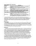
Table 2. Algorithms used in dermoscopy
                Type of algorithm                              Description
                ABCD rule           lesion asymmetry, border, color, differential structure
                A(A)BCD             lesion asymmetry, (differential structures in ≥1 axis), border, color, differential
                                    structure
                ABCDE               lesion asymmetry, border, color, differential structure, elevation
                A(A)BCDE            lesion asymmetry, (differential structures in ≥1 axis), border, color, differential
                                    structure, elevation
                7FFM                7 features of melanoma: pseudopods, radial streaming, regression-erythema,
                                    gray-blue veil, non-homogeneity, irregular pigment network, sharp margin
                Pattern analysis    specific patterns, colors, intensities of pigmentation, configuration, regularity,
                                    characteristics of margin and surface of pigmented lesions
                3-point checklist score  asymmetry of color/structure, atypical network, blue-white structures
                7 point-checklist score  atypical pigment network, blue-whitish veil, atypical vascular pattern, irregular
                                    streaks, irregular pigmentation, irregular dots and globules, regression
                                    structures
                Menzies score       not present: symmetry and single color; at least one feature: blue-white veil,
                                    brown dots, pseudopods, radial streaming, scar-like depigmentation, peripheral
                                    black dots/globules, 5-6 colors, blue/gray dots, broadened network

               Training to increase accuracy. Seven studies analyzed pre-post training in the use of
               dermoscopy to increase the accuracy of detection of melanoma. Most training programs were
               relatively short in duration (1 day to 2 weeks (1 hour per day for 2 weeks in a Web-based
               course)) and consisted of didactic sessions and/or interactive sessions with experienced
               instructors.

               FDA Status
                                                                                            ®
                   The following devices have received Class I FDA approval status: EpiScope  Skin Surface
               Microscope (Model 47300) [Welch Allyn, USA; decision year 1992], NevoScope (TransLite
               USA; decision year 1996), Dermascope (American Diagnostic Corp, USA; decision year 1999),
               and MoleMax (Derma Medical Systems; decision year 1999). The following is a Class II device:
                            ®
               microDERM  (Visiomed AG, USA; decision year 2004).
               Summary
                   Of the 431 abstracts reviewed in this brief, only three were RCTs. Almost all of the primary
               studies on dermoscopy were non-randomized. The non-randomized studies tended to focus on
               features of dermoscopic image that would be of diagnostic interest; digital dermoscopy and the
               use of computer-based analyses; and evaluations of different algorithms and classification
               schemes. We did not identify any controlled studies examining the use of dermoscopy to
               increase the detection rate of early stage melanoma. The primary studies that reported patient
               outcomes largely focused on number of new lesions and how lesions had evolved. No study
               reported on how the addition of dermoscopy affected survival from melanoma.
                   One RCT did compare dermoscopic evaluation and naked-eye examination in 73 primary
               care physicians in Italy and Spain and inferred the effect of the addition of dermoscopy on the
               likelihood that a primary care physician would fail to refer a patient with suspicious skin lesions
               for a second expert opinion. A second RCT of 913 patients in Italy examined the downstream
               effect on the number of skin lesion excised for diagnostic verification with the addition of
               dermoscopy in a pigmented lesion clinic.


                                                             21